
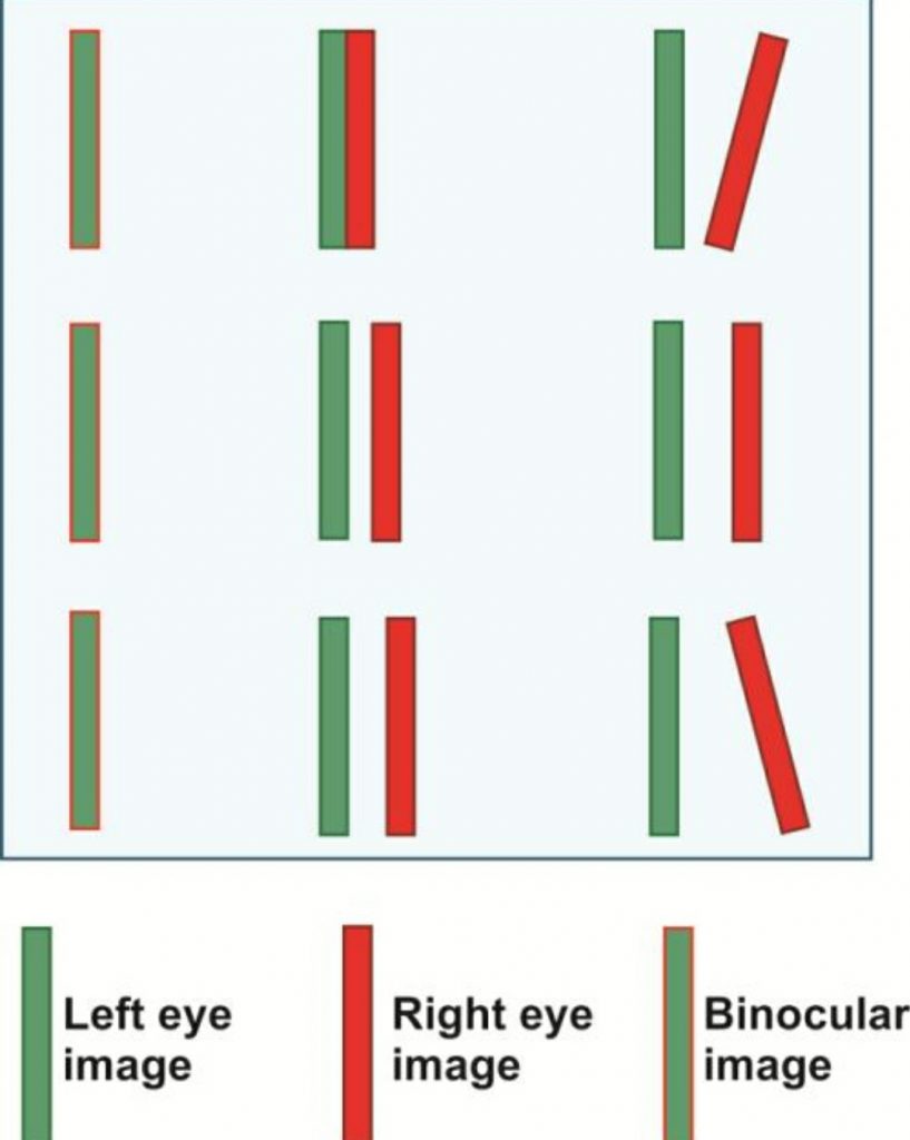
Diplopia refers to the simultaneous perception of two images of a single object that may be displaced horizontally or vertically in relation to each other. It is also called double vision. The condition can affect balance, movement, and reading ability.
It is of two types – (1)Physiological, (2)Pathological
Physiological Diplopia Or “normal double vision” is a phenomenon where objects are doubled, either in front of Or behind, whatever target you are focusing on. Usually, it doesn’t impinge on consciousness.
Pathological Diplopia divided into two types
(a)Binocular Diplopia, (b) Uniocular/ Monocular Diplopia.
In monocular or uniocular Diplopia an object appears double even when only one remains open. This is much less often the case than Binocular Diplopia. This occurs when the two images of one object form on the two different parts of the retina. Monocular Diplopia can occur to the following reasons-
- Dry eye
- Subluxated clear lens
- Large peripheral iridectomy, iridodialysis Or polycoria
- Lens opacity or irregularitiy
- Retinal abnormalities
- Keratoconus
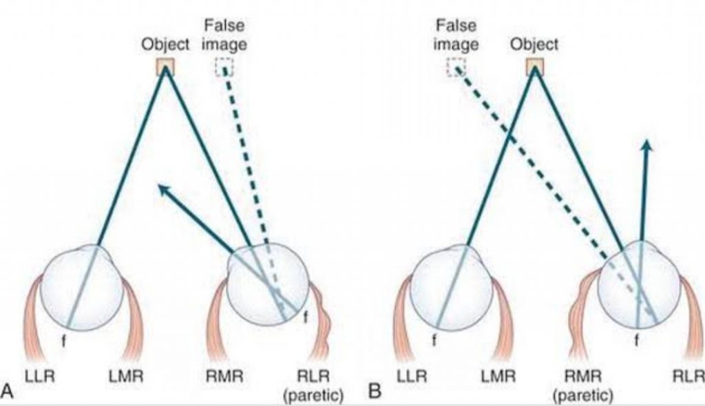
In Binocular Diplopia an object appears double with both eyes remaining open. Here, the image of an object falls on the fovea of one eye and on the extrafoveal area of the opposite eye. The causes of Binocular Diplopia are –
- Paralysis of Extraocular Muscles
- Displacement of the eyeball
- Deviation of rays of light in one eye
- Mechanical restriction of the movements of the globe, pterygium, symblepharon, thyroid ophthalmopathy etc.
- Disparity of image size between two eyes as occurs in acquired high anisotropia
Diplopia has one true image and the other image is false. Binocular Diplopia disappears when one eye is closed. It is of two types
(I) Crossed Diplopia and (II) Uncrossed Diplopia
Crossed Diplopia, which is also called Unharmonious/ heteronymous Diplopia because in this Diplopia the false image is seen on the opposite side. It occurs in divergent squint as in mediak rectus paralysis.
Uncrossed Diplopia, which is also called hamonious Or homonymous because in this Diplopia the false image is on the same side as deviation. It occurs in convergent squint as in lateral rectus paralysis.
For children, diagnose of Diplopia is very difficult because children can’t express what they see. In this case, we must be followed that –
- he/she covering his/her hand to see
- he/she turning his/her head in an unusual way
- he/she narrowing his/her eyes to see
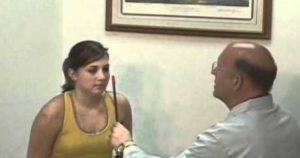
Examination of Diplopia are
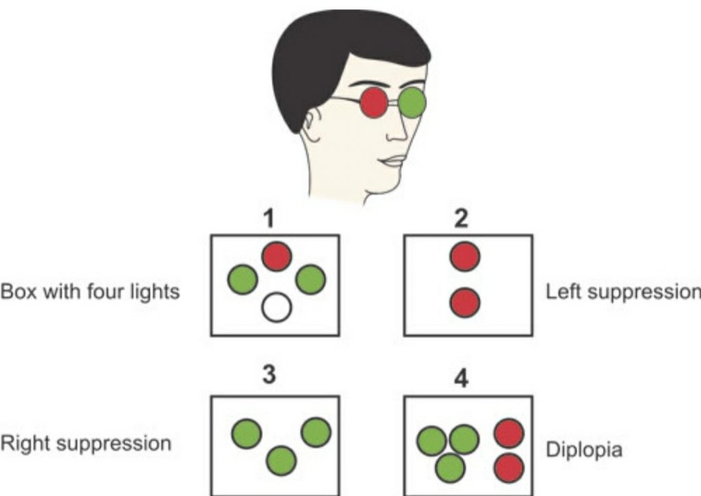
• Worth’s four-dot test – In Diplopia, patient sees five lights ( 2red and 3 green) .
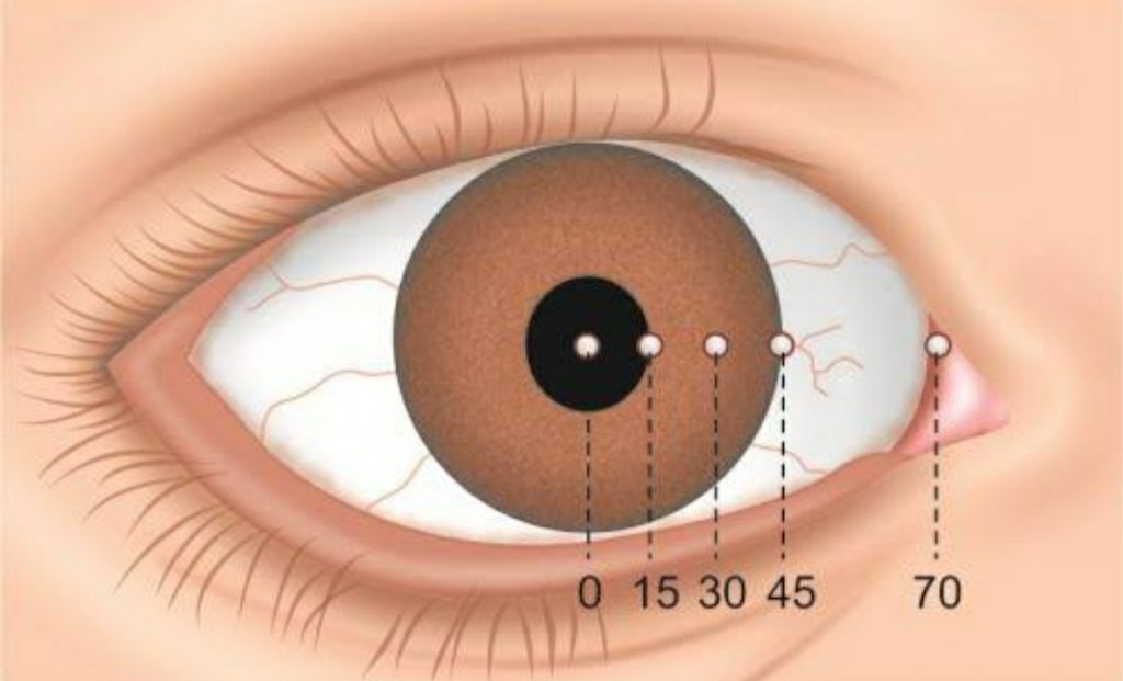
• Hirschberg corneal light reflex test – to measure the degree of Diplopia.
•Cover test
• Diplopia charting
For Treatment
• We must have to find out the cause and then treat on it.
• Occlusion therapy
• Refraction by Prism
• Eye exercise
• Opaque contact lens.


