
Introduction – Binocular single vision is the condition in which two eyes act together. so that the two dissimilar objects came in each eye and the presence of fusion is the blending of sight from the two eyes to a single percept.

Advantages of binocular vision1. Enlargement of the field of vision .2. Help to detect depth perception.3. Binocular vision is a single vision. 4. Compensation for the blind spot.
Grades of binocular single vision
1. Simultaneous macular perception2. Fusion3. Stereopsisthere is one more deviation for fusion that is sensory fusion and motor fusion.
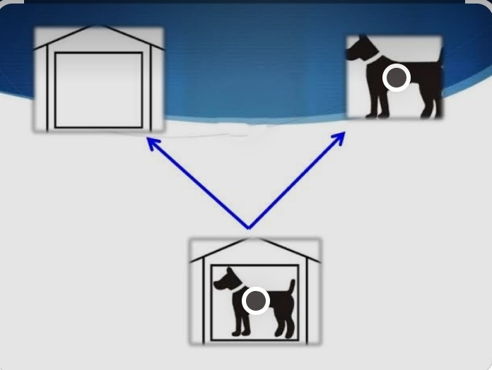
Simultaneous macular perception- It is the procedure in which simultaneously antagonist two objects are superimposed from a complete picture.
Example- one eye saw a hut and the other eye saw a dog and in the presence of simultaneous macular perception both eyes saw a dog in a hut.

Fusion-It is the procedure in which two similar but incomplete objects are superimposed from a complete picture. For example- one eye saw a fox wearing blue pants and the other eye saw a fox wearing a red shirt. And the presence of fusion in both eyes saw a fox wear a red shirt and blue pants.

Sensory fusion – the images not only must be located on corresponding retinal areas but also must be sufficiently similar in size, brightness and sharpness to permit sensory. It’s call sensory fusion.
Motor fusion – It is the ability to align the eyes in such a manner that sensory fusion can be maintained. The stimulus for these fusional eye movements is retinal disparity outside panum’s area and the eyes moving in the opposite directions.
Stereopsis- It is the ability of the eyes to depth perception.

Tests for binocular single vision Test for retinal correspondence–1. Bagalini’s striated glasses test2. Worth’s 4 dot test 3. After image test 4. Red filter testTests for suppression–1. Worth’s 4 dot test 2. 4∆ prism base out test 3. Red filter testTests for fusion–1.worth’s 4 dot test2. Bagalini’s striated glasses testTests for stereopsis–1. Lang’s 2 pencil test2. Random dot test3. TNO test 4. Lang’s stereo test
Worth’s 4 dot test This is a dissociation test that can be used with both distance and near.
Procedure- – The patient wears a red glass in front of the right eye.- And a green glass in front of the left eye- The patient then views a box. There are four lights one red, two green, one white.

Results- – If BSV is present patient sees all four lights.- If the manifest deviation is present patient sees all four lights there is also present ARC.- If the patient sees two red lights there is left suppression present.- If the patient sees three green lights there is right suppression present.- If the patient saw two red lights and three green lights together there is diplopia present.- If the patient sees two red lights and three green lights alternatively there is alternative suppression present.

Bagolini striated glasses
This is the test to detect BSV, ARC, and suppression.
Procedure– The two lenses are placed at 45° and 135° in front of each eye and the patient fixes a small light source.- Each eye perceives an oblique light.- Dissimilar images are thus presented to each eye under binocular viewing conditions.
The result– If the patient sees two streaks intersect at their centers in the form of an oblique cross there is presence of BSV or ARC in the presence of manifest strabismus.- If the patient sees two lines but they do not form a cross there is diplopia present.– If the patient sees one streak there is no simultaneous perception and suppression present.- If the patient sees a small gap in one of the streaks there is central suppression present.

After image test, This test is performed by synoptophore.
Procedure– RE is stimulated by an illuminated horizontal line for 10-15 sec and the patient asks to see at its center.– LE is stimulated with an illuminated horizontal line for 10-15 sec.– The patient is asked to open and close his/her eyes after a few sec . Then look at the wall.
Result– If the patient has seen a perfect cross there is present NRC.- In ARC with ET patient saw the horizontal line in front of RE and the vertical line in front of LE.- In ARC with XC patient saw the vertical line in RE and the horizontal line in front of LE.

4∆ prism base out test This test has been used to confirm the presence of a monofixation syndrome.
Procedure- The patient fixed to a small light then 4∆ bases out prism placed in front of one eye. In a normal patient, this test shows two movements. One version movent and another fusional convergence movement.
Result- – when the prism is placed in front of a dominant eye then both eyes show version movement.- when the prism is placed in front of the non-dominant eye there is no movent in both eyes.

Red filter test this test is applied for diplopia.
Procedure- the red glass was placed in front of the patient’s eye and the patient asked to see a light source with the other eye.
The result– In the case of orthophoria patient will see a single red light.- In the case of esotropia patient will see one red and one yellow light. There is diplopia uncrossed.- In the case of exotropia patient will see one red and one yellow light. There is diplopia crossed.

Anomalies of binocular single vision1. Suppression.2. Amblyopia.3. Abnormal retinal correspondance.4. Confusion.5. Diplopia.
Suppression-It is a cortical phenomenon characterized by a decreased sensitivity to visual information from one eye under binocular conditions.
Amblyopia-In this condition loss of vision in one or both eyes for which no cause can be found by the physical examination of the eye.Types-
1. Strabismic amblyopia 2. Stimulus deprivation amblyopia 3. Isoametropia amblyopia 4. Anisometropic amblyopia 5. Meridional amblyopia treatment- 1. Occlusion therapy 2. Penalization 3. Pharmacologic manipulation
Abnormal retinal correspondence-When the fovea of one eye has a common visual direction with an extrafoveal area in the other eye. This is generally seen if the angle of the squint is small and the extrafoveal point is close to the fovea. It’s call ARC.
Confusion-When corresponding retinal areas are stimulated by dissimilar stimuli and two different objects are seen at one point in the field that creat confusion in our eye.
Diplopia-Simultaneous perception of two images of one object this condition is called diplopia.Types- 1. Binocular diplopia 2. Uniocular diplopia


