
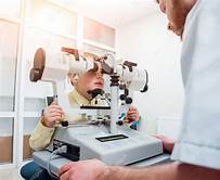
It is a useful orthoptics tool specifically for the diagnosing the subjective and objective angles of squint. This is the instrument that performs a comprehensive assessment of binocular vision by training the ocular muscles, allowing the visual stimuli to be projected on each eye separately.
•What parts does it have?
The synoptophore consists of its base, a metope, a patient chin rest, a volume and frequency control buttons of the illuminating sources, two tubes each 15.5 cm in length, with a + 6.50D collimating lens at each end and a socket for the other end transparencies.
It stands on a cast iron base with one movable foot adjustable by a screw. The base are attached gun metal columns which carry the illuminating systems, slide holders, reflecting mirror and lenses. Each slide holder can be adjusted for vertical and torsional deviations by separate screws.
•Optical principles of synoptophore-
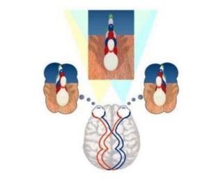
The basic principle of the instrument is the mechanical separation of vision into the right and left eye.
Haploscopic principles- Synoptophore are designed on the principles of division of ‘physical space’ into two separate areas of ‘visual space’ each of which is visible to one eye only.
•How it is performed-
The +6.50 D eye pieces consists of collimating lenses, to completely relax the accommodation. The inter-pupillary distance is adjustable. The tubes can be moved from a convergence position of 70° to a divergence position of 40° . Each tubes can be locked in desires positions and when both are rocked from side to side for moved in a converging or diverging position to give, besides the negative and positive convergence, the range of fusion. The illuminating system is very constructed to avoid any intense reflection and there are rheostats provided which can regulate the intensity of the light. This arrangement results in a rapid flashing of the light to stimulate the under-functional macula.
At the end of each limb there are different sets of slide inserted in slide carriers which can be seen with the two eyes. When the mirrors are placed at 45° to the direction of the tubes, the pictures are fused. Horizontal, vertical and rotatory movements are controlled and measured in scales. Centrally there is a chin and forehead rest.
In new instruments Haidinger’s brushes and after image devices in the two arms of synoptophore have been incorporated which are useful for diagnosis and treatment.
•Uses of synoptophore-
I) Measurements of deviation: Synoptophore measures both objective and subjective angle of squint. All types of heterophorias and heterotropias can be measured accurately with it.
II)Tests for sensory function- it includes,
A) Estimation of grades of binocular vision: Worth has described three grades of binocular single vision, which are best tested with the help of synoptophore. These are,
a. Measurements of simultaneous macular perception that is 1st grade of binocular single vision. This is achieved by placing two different slides like bird and cage in the slot of the instrument, then patient focused through the tubes, if there is simultaneous perception patient will see the bird in the cage.
b. Measurements of sensory and motor fusion that is 2nd grade of binocular single vision. This is achieved by two incomplete but similar images placing two slides, if there is fusion patient will see one complete image.
c. Measurements of stereopsis that is 3rd grade of binocular single vision. It consists of ability to perceive the third dimension or depth perception.
B) Detection of normal (NRC) / abnormal retinal correspondence (ARC) – In normal retinal correspondence (NRC), subjective and objective angle of squint are equal. In ARC, objective angle is greater than the subjective angle.
III) Measurements of primary and secondary deviations and also deviation of 9 gaze of position.
IV) Test to detect suppression. It is a temporary active cortical inhibition of the image of an object formed on the retina of the squinting eye. When the fixating eye is covered, the squinting eye fixes, suppression disappear. This phenomenon is detected by synoptophore test.
V) Measurements of accommodative convergence to accommodation ratio (AC/A).
VI) Measurements of torsion.
VII) Measurement of fusional reserve: the fusional vergences available in excess of the amount necessary to overcome the phoria and to bring the eyes into orthoposition is called relative vergences or fusional reserve. This can be measured by synoptophore.


