In a state of normal binocular single vision, there exists a precise physiological relationship between the corresponding points of the two retina. Thus, the foveae of two eyes act as corresponding points and have the same visual direction is adjustment is called normal retinal correspondence (N RC). When squint develops, patient may experience either diplopia or confusion. To avoid these, sometimes there occurs an active cortical adjustment in the directional values of the two retina. In this state fovea of the normal eye and an extrafoveal point on the retina of the squinting eye acquire a common visual direction, i.e. become corresponding points. This condition is called abnormal retinal correspondence
Signs & symptoms:
. Eye turn
. Absence of diplopia (seeing two images of the same thing)
. Avoidance of eye contact
. Avoidance of visually demanding tasks
. Better than expected performance on tasks requiring binocular vision
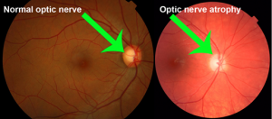
. Post-surgical increase in the angle of deviation
Types:
The type of ARC depends on the objective and subjective angles of anomaly. The objective angle is the motor deviation or the angle of deviation of the eye that is seen. The subjective angle is the sensory deviation or how far off the extra-foveal retinal point is from the actual fovea.
There are three different types of ARC: harmonious unharmonious paradoxical.
In harmonious ARC, the objective angle is equal to the subjective angle.
In Unharmonious ARC, the subjective angle is less than the objective angle.
If the localization of the subjective and objective angles is crossed or uncrossed it is called paradoxical ARC.
Tests to detect abnormal retinal correspondence:
- Worth’s four dot test
- The Bagolini striatrd glass test
- Cover test
- Synoptophore
- Red filter test
- After image test
The Worth 4-dot test can assess binocular vision can evaluate for peripheral sensory fusion and suppression of the fovea. In NRC the patient would see all four dots and in ARC the patient would be diplopic with the deviation adjustable by prism.
The Bagolini striated glasses have no power to them, and the visual acuity of the patient is unchanged. The lenses are placed at right angles at 135 degrees in the right eye and 45 degrees in the left eye with narrow striations that run parallel to each other. Seeing the lights cross as an X means that the patient has normal vision and NRC. Therefore, if it is known that the patient has strabismus and they also report an X – this would indicate ARC.
The cover test can be used to detect the presence and qualities of strabismus that is always present. The occluder is held in front of and then removed from each eye at a time. When the preferred eye is covered, the non-preferred eye may be deviated and will adjust to fixate indicating a tropia. When the non-preferred eye is covered, the preferred eye should not move. Further testing for ARC is warranted if misalignment with normal vision is seen.
Testing with a synoptophore can be used to assess the subjective and objective angle of deviation and any anomalies in binocular vision when an image is presented to each eye. A difference between the subjective and objective angles (the angle of anomaly) indicates that the patient has a form of ARC.
The red filter test can be used to unmask a diplopia in a patient, as well as suppression and ARC. A red filter is placed over the patient’s eye and the patient is asked to focus on the white circle. If an esotropic patient reports crossed diplopia, or an exotropic patient reports uncrossed diplopia, this could indicate ARC. If the diplopia remains with use of a prism, this could also indicate ARC.
The after image test can also assess for ARC and can directly ascertain the foveal direction of the eye that is offset. A vertical light is beamed in one eye, and a horizontal light is beamed into the other eye. If the patient has NRC they will see an after image that looks like a cross, and if the patient has ARC with a known strabismus they will also see a cross.
Treatment:
ARC requires a vision therapy program. While vision therapy may not totally cure ARC, it may substantially improve visual function and quality of life.
A vision therapy program trains the eyes and brain to work together more efficiently and quickly. It corrects certain eye alignment and other binocular vision problems.
Vision therapy includes specific eye exercises paired with special lenses, prisms or eye patches to help improve poor visual skills and binocular vision problems. The program is designed and supervised by your eye doctor.
To learn more or find out how you can strengthen your or your child’s visual skills, schedule an eye evaluation with Visual Symptoms Treatment Center today.
")