
INTRODUCTION
When visual function develops normally, individual posses a binocular single vision, means a point-to-point normal correspondence of two retinae. But in strabismus, the alignment of the corresponding retinal point is disturbed. Thus, the fixation point is imaged in the center of the fovea of the non-deviating eye and some extrafoveal (peripheral retinal) point in the deviating eye. This results in diplopia and confusion.
Diplopia – Diplopia occurs due to formation of image on dissimilar points of two retinae.
Confusion – Confusion occurs due to formation of image of two different objects on the corresponding points of the two retinae.
SENSORY ADAPTATION
A sensory adaptation may be defined as the manner in which a patient makes sensory adjustments to an interruption in the normal binocular single vision caused by the occurrence of squint.
There are three mechanism of sensory adaptation.
- Suppression
- Amblyopia
- Anomalous retinal correspondence.
SUPPRESSION
It is define as active central inhibition of the desperate and confusing image originated from the deviated eye.
TYPES OF SUPPRESSION
- Depending upon the etiopathogenesis
- Physiological suppression
- It refers to the suppression present in everyday life of every individual having normal binocular vision.
- It occurs to avoid retinal rivalry and physiological diplopia.
- It is very helpful in everyday life, as it allows greater concentration on the object of interest.
- For example – While using monocular microscope, monocular telescope.
- Pathological suppression
- It refers to the suppression of the image of one eye that occurs to avoid diplopia and confusion.
- It occurs in patient with strabismus and anisometropia.
- Facultative – It occurs under normal binocular vision but ceases when dominant eye is covered.
- Obligatory – It occurs under all condition and remain even when the dominant eye is covered. This type of suppression leads to amblyopia and decrease in vision in effected eye.
- Depending upon the retinal area where image is suppressed
- Foveal suppression
- Macular suppression
- Peripheral suppression
- Depending upon the constancy
- Intermittent
- Constant
- Depending upon the eye involved
- Monocular
- Alternating
MECHANISM OF SUPPRESSION
Retinal rivalry is a situation that occurs when dissimilar objects are presented to the two eye. Thus struggle for dominance of each eye is considered a prerequisite to suppression.
The extent of the area of the retina that becomes suppressed and the time period during which suppression occurs depends upon the content of the visual field.
Suppression does not occur over the whole retina. It is restricted or localized to a region around fovea of the deviating eye.
Electrophysiological studies give normal ERG and reduced amplitude of VER in patient with suppression.
TEST FOR SUPPRESSION
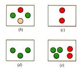
- Worth four dot test
To diagnose the suppression involving the peripheral retina.
For this test red-green goggles are used. Red colour in right eye and green colour in left eye.
The patient having left suppression will see only two red lights and that having right suppression will see only three green lights. In the presence of alternate suppression patient will see alternately two red lights and
three green lights.
- 4D base out prism test
- This test is performed for detection of small angle heterotropias and presence of central suppression scotoma.
- To perform this test patient fixates a penlight, a 4D prism with base out is placed in front of right eye and examiner observes the presence of biphasic corrective movement of left eye.
- This is absent in presence of central suppression scotoma.
MECHANISM OF BIPHASIC CORRECTIVE MOVEMENTS
The prism displaces the image from the fovea of right eye towards a point on a temporal half of the retina.
The relaxation movement of right eye will elicit conjugate movement of both eye of left side.
If right eye has no Foveal suppression this displaces the image in the left eye form the fovea to temporal retina. Thus left eye will make fusional movement in opposite direction if no foveal suppression is present.
- Diplopia test or red glass test
Patient is asked to fixate a spot light at 6m and a prism bar cover test is carried out with prism bar in front of the fixating eye, till deviation is neutralized.
The prism bar reading at this point is equals to the objective angle of squint.
A red filter is placed then placed in front of the deviating eye and the patient is asked to describe what he/she sees.
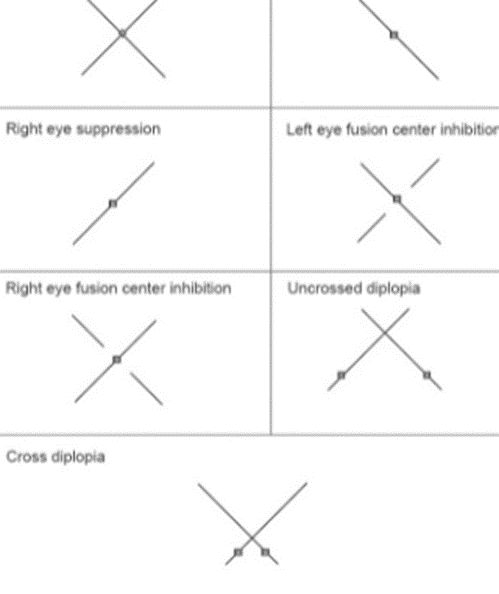
- Bagolini striated glass test
A pair of striated glasses is used in front of each eye.
A source of light is seen as a line at right angles to striations.
Symmetrical cross response in absence of manifest squint, indicates NRC and in presence of manifest squint indicates ARC.
Single line indicates suppression in other eye.
- Visual acuity test with project-O-chart slide of American optical
- Synoptophore test
TREATMENT FOR SUPPRESSION
INDICATION
- Patients with intermittent tropias
- Patient with convergence insufficiency
- Person with constant tropias
- Person with fully corrected accommodative esotropias
- Post surgical patients with slight under correction.
METHOD OF TREATMENT
- Proper refractive correction
- Occlusion therapy
- Alignment of visual axes
- Anti suppression orthoptic exercise
- Diplopia exercises
- Exercise with use of red filter
- Exercise with major amblyoscope
- Exercise with cheirscope
- Exercise with Tibbs binocular trainer.

")


