The retinoscope consists of a light, a condensing lens that concentrates the light, and a mirror. During the procedure, our doctors use the retinoscope to shine light through the pupil, then moves the light vertically and horizontally across each eye and observes how the light reflects off the retina.
The types of the retinoscope then there are two types of retinoscope which are streak and the other one is the spot.Both are used in different retinoiscopy techniques. But if we do a comparison between these two then streak is more useful. Let us talk about both retinoscopes in detail.
Streak retinoscope- The streak retinoscope was first developed by Jack C Copeland. He is also known as the father of streak Retinoscope He patented this in 1927. This produces a linear beam that rotates through the ocular medium. It includes two modes one is Plano mode and the other one is converging mode. In most cases, this streak retinoscope is used with Plano mode. It offers less illumination in comparison to the spot retinoscope. It can be rotated to any axis. It can be changed from convergent to divergent light.
Spot Retinoscope-The first electric retinoscope was developed by Wolff in 1991. This includes a tiny bulb that further helps to spotlight the eye. As this type of retinoscope only has a Plano mode so there is no adjustment needed. In comparison to streak retinoscope, it offers more information. This spot of light can be changed by a slide knob. It can be made larger or smaller in diameter.
The basic principle of retinoscopy is the Foucault test. In this test, a knife edge placed on the principal axis of an optical system (S) intercepts a bundle of rays coming out of (S). Depending on the position of the knife edge, various distributions of light and shadow can be observed on the anterior surface of (S).
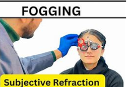
Retinoscopy provides a more accurate result of refractive error in a greater array of patients than autorefraction, although autorefraction is a useful and reliable alternative in many ‘standard’ adult patients and can be particularly accurate at determining astigmatism. Autorefractors should not be used with young children without cycloplegia because of proximal accommodation errors producing significantly more minus results than subjective refraction, particularly in young hyperopes. Retinoscopy also provides a sensitive assessment of the ocular media (e.g. early detection of cataracts, keratoconus), can be used to determine refractive error at distance and near, identify accommodative dysfunction, and is portable, less expensive and less likely to break down.
Retinoscopy’s major disadvantage is that it requires several years of training to become proficient at using it. When a subjective refraction is not possible, limited or unreliable, it is preferable to have more than one assessment of objective refractive correction.
There appears to be no research literature that compares the accuracy of streak or spot retinoscopes or refractions using negative or positive cylinders. The procedure will be described for streak retinoscopy, but spot retinoscopy appears an acceptable alternative. Positive cylinders have the advantage of making retinoscopy easier to learn, as ‘with’ movement is typically easier to see than ‘against’ movement. However, negative cylinders are preferred as they are standard in phoropters. In addition, there is the possibility of stimulating accommodation during subjective refraction when removing a plus cylinder from a trial frame to replace it with one of another power. For these reasons the procedure will be described using negative cylinders.
READ MORE ARTICLE: