
WHAT IS RETINOSCOPY
RETINOSOPY also called as skiascopy or shadow test is an objective method of finding out the error of refraction by the method of neutralization.
PRINCIPAL
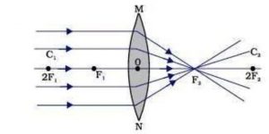
RETINOSCOPY is based on the fact that when light is reflected from a mirror into the eye the direction in which the light will travel across the pupil will depend upon the refractive state of the eye .
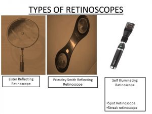
TYPSES OF RETINOSCOPY
- Reflecting mirror retinoscope –
- Plane mirror
- Priestley – smith’s mirror (plane, concave mirrors)
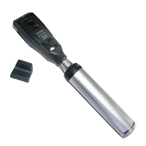
- Self illuminated retinoscope –
- Spot retinoscope
- Streak retinoscope
-
WHAT IS STREAK RETINOSCOPEStreak Retinoscopy is a retinoscope which projects into the patient’s eye an oblong streak which can be adjusted in width and rotated in various meridians .Basic principal in streak retinoscope – there are two systems operating in streak retinoscopy :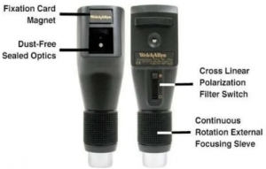
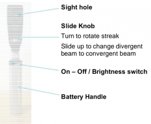
Projection system – it ilumunates the retina and major components such as –
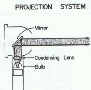
Light source, condensing lens, mirror, focusing sleeve, current source
Observation system – it allows the observer to see the retinal reflex of the patient .
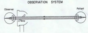
- Light passed from the patients retina
- Light pass through the mirror
- Light go onto the observer’s retina
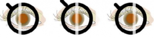
LOOKING AT THE RETINOSCOPIC REFLEX MOVEMENTS
With Movement –
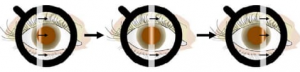
Against Movement –
No Movement (neutral) –
METHOD
- Room lights should be dim.
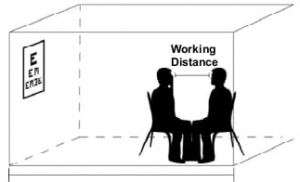
- Ask the person to look at a large fixation target that is at least 3 m away.
- Adjust the trial frame and fit it to the person.
- Sit facing the person so that your line of sight is close to the person’s visual axis. Sit so that:– your head is in front of the person’s right eye– the person can see the fixation target with their left eye
– your eyes are level with those of the person being examined.
- Keep your head straight so that you do not block the person’s view of the fixation target.
- Make sure you are the correct working distance away from the person.
- Blur the eye not being examined with a plus lens – a working lens works well.
- Check that the light beam is divergent .
- Examine the right eye first:
– retinoscope in your right hand
– look through sight hole with your right eye.
- Examine the left eye second:
– retinoscope in your left hand
– look through sight hole with your left eye.
- Keep both of your eyes open.
- Remind the person to look at the fixation target and not at your retinoscope light.
Neutralising astigmatic errors:
- There are two ways to neutralise astigmatic refractive errors:
– using spherical and cylindrical trial lenses
– using spherical trial lenses and an optical cross.
- You only need to use one of these methods.
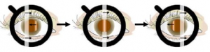
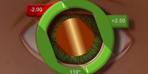
ALIGNMENT OF STREAK RETINOSCOPY REFLEX
The streak of light from a retinoscope can by rotated so that all meridians of an eye can be examined.
Usually the horizontal meridian is scoped first, and then the streak is rotated so that the vertical meridian and oblique meridians are examined.
N.B – • When the streak is vertical (90°)
→ the horizontal (180°) meridian of the eye is being scoped
→ this meridian has an axis of 90° (like the orientation of the streak).
- When the streak is horizontal (180°)
→ the vertical (90°) meridian of the eye is being scoped
→ this meridian has an axis of 180° (like the orientation of the streak).
- When the streak is at an oblique angle
→ the meridian perpendicular to that oblique meridian is being scoped
→ this meridian has an axis that is the same as the orientation of the streak.
Spherical refractive error:
- The ret reflex for a spherical refractive error looks the same in all meridians.
- The same trial lens power will neutralise all meridians.
Astigmatic refractive error:
- The ret reflex for an astigmatic refractive error looks different in different meridians.
- There are two principal meridians that are perpendicular to each other.
- If one of the two principal meridian is being scoped
→ the streak is aligned with the ret reflex
→ we say that we are scoping “on-axis”.
- If other meridians (not the principal meridians) are scoped
→ the streak is not aligned with the ret reflex – there is a “break” in the ret reflex.
→ we say that we are scoping “off-axis”.
- Each principal meridian must be scoped and neutralised individually.
- When we scope a principal meridian the streak orientation is perpendicular to that meridian (but in the same direction as its axis).
Finding principal meridians:
- Sweeping on-axis means that you are scoping one of the principal meridians.
- Sweeping off-axis means that you are not scoping one of the principal meridians.
- To help you scope on axis, look at the ret reflex:
– break ,brightness ,width
ADVANTAGES
- Self illuminated (light source and mirror are incorporated in one )
- Easily manipulated and the intensity and the type of beam can be easily controlled .
DISADVATAGES
- The transparent sealed optics easily have the finger print .
- The light too bright – induces pupil constrict
- The batteries will easily lasting – the bulb is bright
CONCLUSION
As we have seen , retinoscopy is an essential technique in optometry field as it can be very helpful to the patient such as children, nonliterate , hearing impaired , language disorder, or indecisive
Whatever the brand that observer used not determine the accuracy , the endpoint lies in the observer’s successful performance and interpretation of the art.


