
Retinoscopy is also known as skiascopy, shadow test or pupilloscopy or koreoscopy. It was introduced by Bowman in 1859. It is an objective method of finding out the error of refraction by utilizing the technique of neutralization. It is based on the fact that when light is reflected from a mirror into the eye, the direction in which the light will travel across the pupil will depend upon the refractive state of the eye
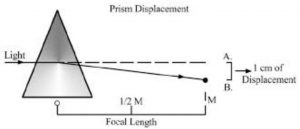
Optics of retinoscopy:- In retinoscopy, an area of the fundus is illuminated by the light reflected into the patient’s eye with the help of a retinoscope. This illuminated area serves as an object and the rays which emanate from this area illuminate the pupillary area and from its image at the far point of the eye.
Prerequisites for retinoscopy:-
- A Darkroom ( Which is preferably 6 m long )
- Trial box ( Which contains spherical and cylindrical lenses of variable plus and minus power and an occluder )
- The trial frame ( preferably of the adjustable type which can be used in children as well as in adults )
- Retinoscope ( two types of retinoscope mirror retinoscope and streak retinoscope )
- A bulb ( Which is used as a source of light, which is required when using a mirror retinoscope )
Uses of cycloplegics in retinoscopy:- Cyclopegics are the drugs that cause paralysis of accommodation and dilate the pupil. These are used for retinoscopy when the examiner suspects that accommodation is abnormally active and will hinder the exact retinoscopy. Such a situation is encountered in young children and hypermetropic. When retinoscopy is performed after instilling cycloplegic drugs, it is termed wet retinoscopy. The commonly employed cycloplegics are as follows –
Atropine:- It is used as a 1 % ointment thrice daily for 3 days before performing retinoscopy. Its effect last for 10-20 days. It is used below the age of 5 years. The period of the post-cyclopegic test is done after 3 weeks of retinoscopy and the tonus allowance is 1 D.
Cyclopentolate:- Cyclopentolate is used as 1 % eye drops in patients between 8 to 20 years of age. One drop of cyclopentolate is instilled after every 10-15 minutes 3 times and retinoscopy is performed about 60-90 min. Its effect lasts for 6 to 18 hours. Tonus allowance is 0.75D.
Homatropine:- Homatropine is a drug it is used as 2% drops. One drop is often instilled every 10 minutes for 6 times and the retinoscopy is performed after 1 to 2 hours. Its effect lasts for 48 to 72 hours. It is used for most hypermetropic individuals between 5 and 25 years of age.
Procedures of retinoscopy:-
The patient is made to sit at a distance of 1m from the examiner. It is even more convenient to sit at arm’s length, i.e. two- thirds meters away.
The patient is normally seated and looking toward the far end of the room.
The Source of light is from behind the patient.
The observer looks through a plane mirror with central perforation, and light is reflected into the patient’s eye.
The mirror is slowly moved from side to side in different meridians and movement of the shadow is noted.
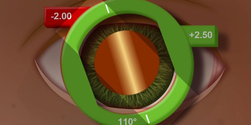
In hypermetropia, emmetropia, and myopia less than 1.00D the reflex moves in the same direction. In myopia of 1.00D, there is no shadow, in myopia of greater than 1 D the shadow moves in the opposite direction.
Increasing convex or concave lenses are placed before the eye until the point of reversal is reached.
Observations of retinoscopy:-
No movement of the red reflex indicates myopia of 1D
With movement of the red reflex along the movement of the retinoscope indicates either emmetropia or hypermetropia or myopia of less than 1D.
Against movement of the red reflex to the movement of the retinoscope implies myopia of more than 1D.
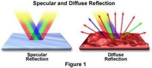
Bright and fast shadow is seen in the pupillary area which moves rapidly with the movement of the mirror, in patients with low degrees of refractive errors.
Dull reflex which moves slowly with the movement of the mirror is seen in patients with high degrees of ametropia.
When using a streak retinoscope of red reflex is narrow with a high degree of ametropia and the wide reflex means a low degree of ametropia.
The calculation for retinoscopy:- When the observer is sitting at 1-meter deduction of the retinoscopic figure is 1 D and 1.50 D deduction when the observer sits at 2/3 rd meter distance, samely 1 D when using atropine, 0.5 D for homatropine and 0.75 D for cyclopentolate
So the refraction of the eye = -1.0D + end point of refraction + 1D/0.75D/0.5D ( when use wet retinoscopy )
Problems in retinoscopy:-
- Red reflex may not be visible or may be poor this may happen with small pupils and hazy media.
- Scissor shadows may sometimes be seen in patients with regular astigmatism with dilated pupils. It is difficult to diminish with the undilated pupil.
- Triangular shadow may be seen in patients with the conical cornea. On moving the mirror, the triangular reflex appears to swirl around its apex.


