Optical Coherence Tomography (OCT) is a non-invasive imaging technology used in various
medical fields, particularly ophthalmology, cardiology, and dermatology, to visualize and
assess biological tissues with high resolution and detail. In this essay, I will delve into the
principles, applications, advancements, and future prospects of OCT, providing a
comprehensive overview of this powerful imaging technique.
Principles of OCT
OCT is based on the principle of low-coherence interferometry, which involves measuring the echo time delay and intensity of light reflected from biological tissues. The key components of an OCT system include a broadband light source, a beam splitter, reference and sample arms, and a photodetector. The light emitted by the source is split into two arms, with one directed towards the sample (tissue) and the other towards a reference mirror. The reflected light from both arms interferes, and the resulting interference pattern is analyses to generate high-resolution cross-sectional images of the tissue.
Types of OCT
Several variations of OCT exist, including Time-Domain OCT (TD-OCT), Fourier-Domain OCT (FD-OCT)FD-OCT is the most commonly used types due to their faster imaging speeds and higher
sensitivity compared to TD-OCT.
Time domain
In time domain OCT the path length of the reference arm is varied in time (the reference mirror is translated longitudinally). A property of low coherence interferometry is that interference, i.e. the series of dark and bright fringes, is only achieved when the path difference lies within the coherence length of the light source. This interference is called autocorrelation in a symmetric interferometer (both arms have the same reflectivity), or cross-correlation in the common case. The envelope of this modulation changes as path length difference is varied, where the peak of the envelope corresponds to path length
matching.
Fourier domain
Fourier-domain (or Frequency-domain) OCT (FD-OCT) has speed and signal-to-noise ratio (SNR) advantages over time-domain OCT (TD-OCT) and has become the standard in the industry since 2006. The idea of using frequency modulation and coherent detection to obtain ranging information was already demonstrated in optical frequency domain reflectometry and laser radar in the 1980s, though the distance resolution and range were much longer than OCT. There are two types of FD-OCT – swept-source OCT (SS-OCT) and spectral-domain OCT (SD-OCT) – both of which acquire spectral interferograms which are then Fourier transformed to obtain an axial scan of reflectance amplitude versus depth. In SS-OCT, the spectral interferogram is acquired sequentially by tuning the wavelength of a laser light source. SD-OCT acquires spectral interferogram simultaneously in a spectrometer. An implementation of SS-OCT was described by the MIT group as early as 1994.
Applications of OCT
1.Ophthalmology:
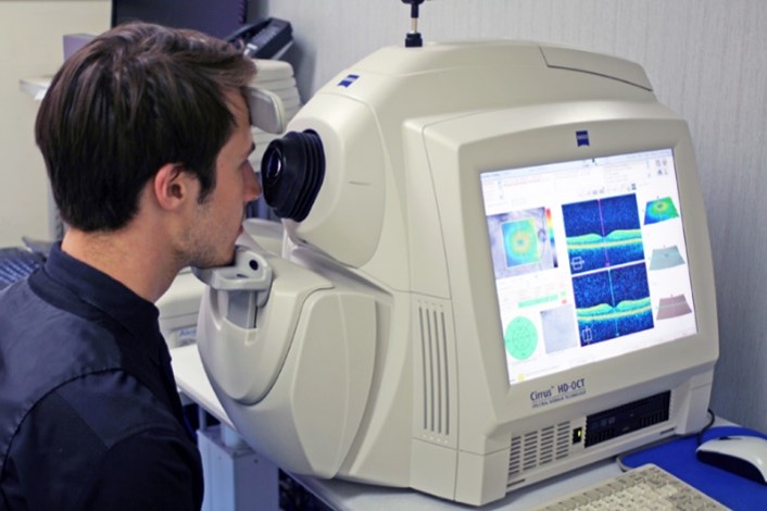
OCT revolutionized the diagnosis and management of various ocular diseases, including macular degeneration, diabetic retinopathy, glaucoma, and retinal vascular disorders. It provides detailed cross-sectional images of the retina, optic nerve, and anterior segment, enabling early detection of pathological changes and monitoring disease progression.
2.Cardiology:
In cardiology, OCT is used to visualize coronary arteries and assess coronary plaque morphology, composition, and vulnerability. It aids in the diagnosis and treatment planning of coronary artery disease and guiding intravascular interventions such as stent placement.
3.Dermatology:
OCT is utilized in dermatology for imaging skin lesions, monitoring wound healing, and assessing skin structure and pathology. It provides valuable information for diagnosing skin cancers, evaluating treatment response, and guiding surgical procedures.
4.Neurology:
OCT is increasingly used in neurology to visualize and quantify structural changes in the central nervous system, particularly in conditions such as multiple sclerosis and optic neuropathies. It helps in monitoring disease progression and evaluating treatment efficacy.
Advancements in OCT
Recent advancements in OCT technology have significantly improved imaging speed, resolution, and depth penetration, expanding its applications and clinical utility. Swept- Source OCT (SS-OCT) and Full-Field OCT (FF-OCT) are emerging techniques that offer enhanced imaging capabilities for specific clinical scenarios. Additionally, developments in image processing algorithms, artificial intelligence, and machine learning have enabled automated segmentation, quantification, and analysis of OCT images, facilitating rapid diagnosis and personalized treatment strategies.
Future Prospects of OCT
The future of OCT holds immense promise, with ongoing research focused on further enhancing imaging performance, expanding clinical applications, and improving accessibility and affordability. Advances in light sources, detection techniques, and miniaturization of OCT systems may lead to the development of portable, point-of-care devices for widespread use in primary care settings and resource-limited environments. Moreover, integration of OCT with other imaging modalities such as fluorescence imaging, confocal microscopy, and photoacoustic imaging holds potential for multimodal imaging approaches that offer complementary information and improve diagnostic accuracy. Optical Coherence
Tomography is a versatile imaging technology with wide-ranging applications in medicine and biomedical research. Its ability to provide high-resolution, real-time, non-invasive imaging of biological tissues has revolutionized the diagnosis, monitoring, and treatment of various diseases across multiple medical specialties. With continued advancements and innovations, OCT is poised to play an even more prominent role in healthcare, enabling earlier detection, personalized treatment approaches, and improved patient outcomes


