Slit lamp is a specialized microscope that employs a focused beam of light to examine the
eyes structures. It consists of a light source and a series of lenses that direct the light in the eye,
enabling healthcare professionals to diagnose and evaluate various ocular conditions. With its ability to
provide a magnified and detailed view of the front and back of the eye, the slit lamp aids in the
assessment of eye health and the identification of issues such as cataracts, glaucoma, corneal defects,
and retinal diseases.
The main parts of a slit lamp typically include:
1. Chinrest: It provides a stable and comfortable platform for the patient's chin and forehead,
ensuring proper alignment and positioning.
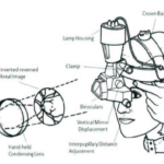
2. Biomicroscope: This is the main body of the slit lamp that houses the optical system,
illuminating system, and observation system.
3. Illumination system: It consists of a light source, typically a halogen, which produces a focused
beam of light. It also includes filters to modify the colour and intensity of the light.
4. Slit beam control: This feature allows adjusting the width, height, angle, and shape of the slit
beam. It helps to focus and direct the light onto specific areas of the eye.
5. Observation system: It includes oculars or binoculars for the examiner to view the magnified
image of the eye. Many modern slit lamps also have a camera or digital imaging system for
capturing images or videos.
6. Filters: Slit lamps have various filters, such as cobalt blue, red-free, or yellow filters, which
enhance the visualization of specific structures or conditions in the eye.
7. Joystick or control handles: These are used to move the slit lamp in different directions, adjust
the height, and fine-tune the focus.
8. Applanation tonometer: Some slit lamps may have an attached tonometer to measure
intraocular pressure by gently flattening the cornea.
Prism use in Slit lamp:-
A prism is sometimes used in slit lamps to assist with certain examinations.
Prisms in slit lamps are primarily used for assessing the eye's alignment and binocular vision. By
placing a prism in front of one eye, the examiner can observe the patient's eye movement and
detect any misalignment or abnormal eye coordination. This is particularly helpful in diagnosing
conditions like strabismus (misaligned eyes) or assessing the patient's ability to merge the
images from both eyes (binocular vision).
The prism can be rotated and adjusted to observe different angles and directions of eye
movement. This assists in measuring the degree of misalignment and determining the
appropriate treatment options.
Overall, the use of prisms in slit lamps enhances the diagnostic capabilities of eye care
professionals and helps them in examining and managing various eye conditions related to
binocular vision and eye alignment.
Mirror use in Slit lamp :
A mirror is commonly used in a slit lamp. It is referred to as a beam splitter mirror. The
beam splitter mirror is located inside the microscope of the slit lamp and its purpose is
to split the light into two pathways: one for observation and one for imaging or
documentation.
The main light source enters the beam splitter mirror and is divided. One portion of the
light continues to illuminate the patient's eye, allowing the examiner to view the eye's
structures through the microscope. The other portion of the light is directed towards a
camera or other imaging device for capturing images or videos of the eye.
The beam splitter mirror is an essential component of the slit lamp, enabling
simultaneous observation and documentation of the eye's examination findings.
Techniques of Slit lamp
There are several techniques used with a slit lamp in order to examine the eye's
anterior segment. Here are some common techniques:
1. Direct Illumination: This is the most basic technique where a narrow beam of light is
directed onto the specific area of interest in the eye. The examiner can vary the size,
shape, and orientation of the slit to focus on different structures.
2. Diffuse Illumination: In this technique, the light is spread out into a wider beam to
provide a more overall view of the eye's structures.. It gives a broader illumination and
helps in assessing the overall health and surface quality of the eye.
3. Indirect Gonioscopy: This technique involves using a special mirrored lens called a
gonioscopy lens to visualize the angle between the cornea and the iris. It helps in the
assessment and management of conditions related to the drainage system of the eye,
such as glaucoma.
