
Indirect ophthalmoscopy is a diagnostic technique used to examine the interior structures of the
eye, including the retina, optic nerve, and blood vessels, by using a condensing lens and a light
source. The examiner holds the condensing lens in front of the patient's eye and uses a bright light
to illuminate the interior of the eye, enabling a magnified view of the retina and other structures.
This method allows for a wide-field view of the fundus, aiding in the detection of various eye
conditions such as diabetic retinopathy, macular degeneration, and retinal detachment. It is an
essential tool in ophthalmic examinations and provides valuable information for the diagnosis and
management of ocular diseases.
WORKING MECHANISM OF INDIRECT OPHTHALMOSCOPY
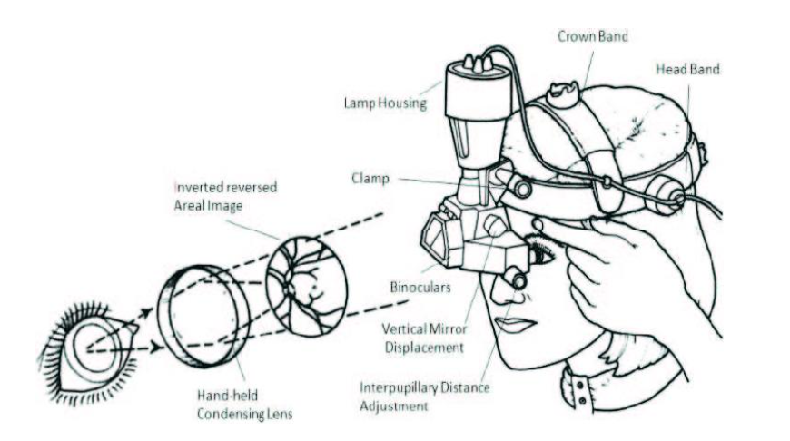
1. Condensing Lens: The examiner holds a condensing lens in front of the patient's eye. This
lens helps to converge the light rays from the fundus (the interior surface of the eye) and
also magnifies the image of the fundus. The type of lens used in indirect ophthalmoscopy is
typically a positive power lens, which aids in focusing light onto the retina and facilitates the
visualization of the structures in the eye.
2. Light Source: An external light source, such as a slit lamp or a handheld ophthalmoscope,
is used to illuminate the interior of the eye. By directing this light into the eye, the examiner
can observe the reflection and transmission of light from the fundus structures.
3. Magnified View: The combination of the condensing lens and the light source results in a
magnified and illuminated view of the fundus. This enables the examiner to visualize the
retina, optic nerve, blood vessels, and other structures in detail.
Overall, indirect ophthalmoscopy allows for a wide-field, three-dimensional view of the
fundus, providing valuable information about the health of the eye and aiding in the
diagnosis of various ocular conditions.
BENEFITS OF INDIRECT OPHTHALMOSCOPY
1. Wide-Field View: It provides a wide-field view of the fundus, enabling the examiner to
visualize a large area of the retina and optic nerve. This is particularly useful for assessing
peripheral retinal pathology and identifying conditions such as retinal holes, tears, or
detachments.
2. Magnified Visualization: The magnification provided by the condensing lens allows for
detailed visualization of fine structures within the eye, such as the macula, optic disc, and
retinal blood vessels. This aids in the detection of abnormalities such as diabetic retinopathy,
macular degeneration, and hypertensive retinopathy.
3. Depth Perception: Indirect ophthalmoscopy allows for a three-dimensional view of the
fundus, providing depth perception that aids in assessing the topography and contour of
retinal lesions or abnormalities.
4. Non-Contact Examination: Unlike direct contact methods, such as contact lens
biomicroscopy, indirect ophthalmoscopy is a non-contact technique. This reduces the risk of
corneal abrasions and makes the examination more comfortable for the patient.
5. Pupil Dilation Not Always Required: In some cases, indirect ophthalmoscopy can be
performed without the need for pupil dilation, making it a more accessible method for
routine screening or urgent eye examinations.
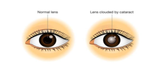
6. Evaluation of Media Opacities: It allows the examiner to visualize the fundus even in the
presence of media opacities, such as cataracts, which may hinder the use of direct
ophthalmoscopy.
In summary, indirect ophthalmoscopy is valuable for comprehensive visualization of the
posterior segment of the eye, aiding in the diagnosis, monitoring, and management of
various retinal and optic nerve conditions.
WHY WE DO INDIRECT OPHTHALMOSCOPY
Indirect ophthalmoscopy is performed for several reasons in the field of ophthalmology and
optometry. Here are some key reasons why this diagnostic technique is used:
1. Comprehensive Eye Examination: Indirect ophthalmoscopy allows for a comprehensive
evaluation of the posterior segment of the eye, including the retina, optic nerve, and
vitreous, providing valuable information about ocular health.
2. Detection of Retinal Abnormalities: It is used to identify and monitor retinal conditions
such as diabetic retinopathy, macular degeneration, retinal detachment, hypertensive
retinopathy, and retinal vascular disorders. The wide-field view and magnified visualization
aid in the early detection of these conditions.
3. Assessment of Optic Nerve Health: Indirect ophthalmoscopy enables the assessment of
the optic nerve head, aiding in the diagnosis and management of conditions such as
glaucoma, optic nerve atrophy, and papilledema.
4. Monitoring Eye Health in Systemic Diseases: It is used to monitor the ocular
manifestations of systemic diseases, such as hypertension, diabetes, and neurological
disorders, which can affect the retina and optic nerve.
5. Evaluation of Traumatic Eye Injuries: Indirect ophthalmoscopy is utilized to assess and
monitor the ocular effects of traumatic injuries, including retinal tears, hemorrhages, and
vitreous disruptions.
6. Routine Eye Examinations: Ophthalmologists and optometrists use indirect
ophthalmoscopy as part of routine eye examinations to screen for retinal and optic nerve
abnormalities, especially in patients with risk factors for ocular diseases.
7. Surgical Planning and Post-operative Monitoring: It is employed in the pre-operative
assessment of patients undergoing ocular surgery, as well as in the post-operative
monitoring of retinal surgeries and other procedures involving the posterior segment of the
eye.
Overall, indirect ophthalmoscopy is a vital tool for the diagnosis, management, and ongoing
care of a wide range of ocular conditions, playing a key role in comprehensive eye care and
disease management.


