
The human eye sight is a highly specialized method to navigate the surroundings. The visual pathway refers to the anatomical structures responsible for the transformation of light energy into electrical action potentials that can be converted by the brain.
Components of visual pathway:
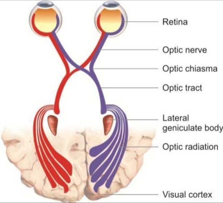
The visual pathway begins at the retina consist of:- •Two optic nerves
• An optic ic tracts
•Two lateral geniculate body
•Two optic radiations
• visual cortex on each side.
•Optic nerve:-
Each optic nerve starts from the optic disc and extends from the ganglion cells of the retina to the chiasma, where the two nerves meet. It contains the afferent fibres of the Pupillary light reflex and some centrifugal fibres.
Morphologically and embryologically the optic nerve is comparable to sensory tract (white matter) of the brain, because of the following points:
•The optic nerve is an outgrowth of the brain.
• unlike peripheral nerves it is not covered by neurilemma (so it does not regenerate when cut).
• The fibres of optic nerve numbering about a million, are very fine, 2-10um in diameter as compared to 20um of the sensory nerve.
•The optic nerve is surrounded by meninges.
•Parts of optic nerve:
The optic nerve is about 47-50mm in length and is anatomically divisible into four parts:-
•Intraocular part [ which transverses the posterior scleral foramen 1mm in length]
•Intraorbital part [which runs in the orbit it is about 25mm in length]
•Intracanalicular part [which transverse the optic foramen, it is about 6-9mm in length]
•Intacranial part [which runs from the optic foramen to the angle of the chiasma and is about 10mm in length]
- Intraocular part: Intraocular part starts from the optic disc as the fibres converge from the retina. They pile up at the margin of the disc more so on the nasal side(optic papila). This shows a central depression from where the vessels emerge. The depression is called the physiological cup of the disc. A dence ciliary structure bridges across the scleral foramen enclosing in its numerous trabeculae the fasiciculi of the optic nerve (lamina cribrosa).
- Intraorbital part: It extends from back of the eyeball to the optic foramen. At the optic foramen it is surrounded by the origin of the ocular muscles, the dural, arachnoid and pial sheaths of the optic nerve which are continuous posteriorly with the meninges of the brain. This part is slightly sinuous to give play for the eye movements. It is closely surrounded by the annulus of zinn and the superior and medial rectus muscles at their origin are closely attached to the dural sheath of the optic nerve. This attachment is responsible for pain on medial and upwards movements of the eyeball in cases of retrobulbar neuritis. The nasociliary nerve, the ophthalmic artery and superior ophthalmic vein cross the nerve superiorly from lateral to medial side. Anteriorly, the nerve is separated from the ocular muscles by the orbital fat.
- Intracanalicular part: This is the third part of the nerve. It is closely related to the ophthalmic artery which lies inferolateral to it and Crosses obliquely over it, as it enters the orbit, to lie on its medial side.
The intracanalicular part of the nerve is 6-9mm in length. The sphenoid and the posterior ethmoidal sinuses lie medial to it and are separated by a thin bony lamina. This relation accounts for retrobulbar neuritis following infection of the sinuses. - Intacranial part: This is the fourth part of the nerve. It is about 10mm long and it runs from the optic foramen to the optic chiasma. It lies at first on disphragma sellae and then on the anterior perforated substance and the medial root of the olfactory tract. The anterior cerebral artery lies above and crosses it from lateral to medial side. The internal carotid artery is at first below and the lateral to the nerve. The ophthalmic artery crosses it inferiorly from lateral to the medial side. The nearer is the nerve to the foramen opticum, the nearer is the artery to the medial border of the nerve.
•Optic chiasma:-
It is a flattened oblong band some 12mm in its transverse diameter and 8mm from before backwards. It is placed at the junction of anterior wall and floor of the third ventricle itself forming the floor of the recess which reaches almost to its anterior border. clothed in pia it lies obliquely with its posterior border higher than anterior part of the cisterna interpeduncularis over the diaphragma sellae, above and behind the so called optic groove on the sphenoid bone. It is thus suspended in and surrounded by cerebrospinal fluid except at its posterior border. On entering the cranium the optic nerve converges towards its fellow of the opposite side intimately clothed in pia mater. The two together from chiasma in front is the anterior cerebral artery and laterally it is related to the internal carotid artery. It rests behind the olivary eminence and is covered by the pia mater in the cisterna basilis of subarachnoid space.
The chiasma is not in contact with the diaphragma sellae, but is separated from it by about 5-10mm so that cisterna basalis lies deep to the chiasma. From its under surface the infundibulum or the pituitary stalk passes downwards and forwards through a hole in the posterior part of diaphragma sellae to be attached to the posterior lobe of the pituitary body.
•Optic tract:-
The chiasma ends into two optic tracts. There are cylindrical bundles of nerve fibers running outwards and backwards from the posterolateral aspect of the optic chiasma. Each tract takes the from of a round band. They run at first between the tuber cinereum and the anterior perforated substance. They continue posteriorly to sweep around the peduncles in association with posterior cerebral artery. Each optic tract consists of fibres from the temporal half of the retina of the same eye and the nasal half of the opposite eye. Posteriorly, each optic tract ends in the lateral geniculate body. The pupillary reflex fibres pass on the pretectal nucleus in the midbrain through the superior brachium. Some fibres terminate in the superior colliculus.
•Lateral geniculate bodies:-
There are two lateral geniculate bodies are oval structures situated at the posterior termination of the optic tracts. Each lateral geniculate body consists of six layers of neurons grey matter alternating with white matter fromed by optic fibres. The fibres of second order neurons coming via optic tracts relay in these third order neurons travels by way of the optic radiation to the occipital cortex (visual center).
•Optic radiation:-
The optic radiations or geniculocalcarine pathway extends from the lateral geniculate body to the visual cortex. They pass forwards and then laterally through the area of wernick as optic peduncles anterior to the lateral ventricle and traversing the retrolenticular part of internal capsule, behind the sensory fibres and medial to auditory tract.
The fibres of optic radiations then spread out fanwise to form a medullary optic lamina. This is at first vertical but becomes horizontal near the visual cortex.
•Visual cortex:-
It is located on the medial aspect of the occipital lobe in and near the calcarine fissure. It is subdivision into the visuosensory area that receives the fibres of the radiations, and the surrounding visuopsychic area.
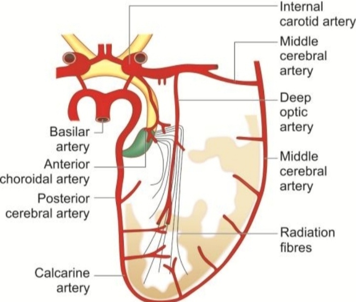
•Blood supply of the visual pathway:-
All the arteries which eventually supply the nerve tissue do so through the pial network of vessels. The visual pathway is mainly supplied by pial network of vessels except the orbital part of the optic nerve which is also supplied by an axial system derived from the central artery of retina. This pial network is rich and fine and extends to the back of the globe. The pial plexus around different parts of the visual pathway gets contribution from different arteries.
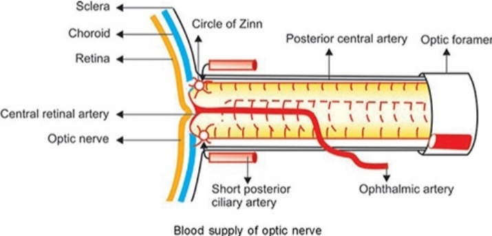
Blood supply of the optic nerve head:-
Needs special mention optic nerve head can be divided into 4 parts-
1. Surface layer
2. Prelaminar region
3.lamina cribrosa
4. Retrolaminar part
- Surface layer of the optic disc is supplied by capillaries derived from the retinal arterioles.
- Prelaminar region is mainly supplied by centripetal branches of the peripapillary choroid with some contribution from the vessels of lamina cribrosa.
3.Lamina cribrosa is supplied by branches from the posterior ciliary arteries and arterial circle of zinn.
4.Retrolaminar part of the optic nerve is supplied by centrifugal branches from central retinal artery and centripetal branches from pial plexus formed by branches from the choroidal arteries, circle of zinn, central retinal artery and ophthalmic artery.


