
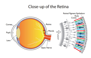
The retina is a layer of photoreceptors cells and glial cells within the eye that captures incoming photons and transmits them along neuronal pathways as both electrical and chemical signals for the brain to perceive a visual picture. The retina is located in the posterior segment and forms the innermost boundary among the other major layers of the eye that include the vascular choroid and the fibrous sclera. Disease manifestations can occur in the retina at different stages of life, many of which severely compromise visual ability and consequently the quality of life.
Structure and Function
The following topics relating to the structure and function of the retina appear below:
- Photoreceptor cells
- Layers of the retina
- Macula
Photoreceptor Cells
Photoreceptor cells include rods and cones and are uniquely located towards the posterior aspect of the retinal sublayers, further away from the pupil where light enters the eye. Rods are more sensitive in dim light (scotopic vision) and reside in the periphery of the retina. Cones are more sensitive in daylight (photopic vision) and capture wavelengths of coloured light. Cones localize in the centre of the retina at the fovea. There are approximately 6 million cones and often more than 100 million rods within the retina. There exist three types of cones including tritons, deuterons, and protons, named for detecting short, medium, and long wavelengths, respectively. In terms of sensing coloured light, each type of cone cell can respectively characterize as detecting blue, green, and red wavelengths. The overlap of detectable wavelength spectrums between the three types of cones results in the visible light spectrum perceived by humans. Rod cells contain rhodopsin, which is a light-sensitive pigment made of retinal that allows for the absorption of photons. Retinal is vitamin A aldehyde, making vitamin A an essential dietary component for the facilitation of the phototransduction pathway. Vitamin A deficiency is a significant risk factor for blindness in young children and remains prominent in under-developed regions, including South Asia and sub-Saharan Africa.
Layers of the Retina
The retina, more specifically, subdivides into ten distinct layers that are described in order from the innermost layers closer to the pupil to the layers further towards the posterior and periphery of the eyeball:
Inner Limiting Membrane – the innermost layer of the retina that forms a smooth boundary against the vitreous humour which fills the vitreous chamber of the eye. The peripheral boundary of this layer consists of Müller glial cells, which function to maintain retinal homeostasis by upholding retinal lamination and by supporting other cells.
Retinal Nerve Fibre Layer – the layer composed of retinal ganglion cell axons mixed with astrocytes and the processes of the Muller cells. The inner limiting membrane serves as the basal lamina for the cells of the retinal nerve fibre layer.
Ganglion Cell Layer – the layer of ganglion cell bodies that project their axons, eventually to form the optic nerve.
Inner Plexiform Layer – this layer is where the axons of bipolar cells synapse onto the ganglion cells. The dendrites of amacrine cells also synapse at this zone and function in modulating the electrical conduction between the bipolar cells and ganglion cells, preventing lateral potentiation.
Inner Nuclear Layer – the layer composed of the cell bodies of bipolar cells, horizontal cells, and amacrine cells. Bipolar cells function as channels that transmit and encode various synaptic inputs from photoreceptor cells onto ganglion cells. Horizontal cells provide feedback modulation onto rod and cone cells.
Outer Plexiform Layer – the region where projections from photoreceptor cells synapse with the dendrites of the cells residing in the inner nuclear layer.
Outer Nuclear Layer – the layer containing the cell bodies of both rods and cones.
External Limiting Membrane – the region that is composed of gap-junctions between photoreceptor cells and Muller cells; it separates the cell bodies of the rods and cones from their inner segments and outer segments.
Photoreceptor Layer – the region consisting of the inner segments and outer segments of rods and cones. The outer photoreceptor segments consist of membrane-bound discs that contain the light-sensitive pigments such as rhodopsin that are necessary for phototransduction. The inner segments house the abundance of mitochondria needed to meet the high metabolic demands of the photoreceptor cells.
Retinal Pigment Epithelium – the outermost retinal layer that spans a width of a single cell located between the neural retina and the Bruch membrane, adjacent to the highly-vascularized choroid layer. The retinal pigment epithelium (RPE) contributes to the blood-retinal barrier in conjunction with the endothelium of the retinal vessels and has many functions including ion and water transport and secretion of growth factors and cytokines. The RPE cells intermingle with the outer segments of the rods and cones. This proximity allows for the recycling of all-trans-retinal back into 11-cis-retinal and its delivery back to the cones and rods to be used again for phototransduction.RPE cells are crucial in the support and maintenance of both photoreceptor cells and the underlying capillary endothelium.
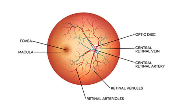
Macula
The macula, also called the macula lutea for its yellowish pigmented appearance, makes up the most sensitive area of the retina, offering the highest visual acuity. It is found temporally from the optic disc upon fundoscopic examination. Lutein and zeaxanthin are carotenoids that make up the macular pigments and produce the yellow colouring. These macular pigments are known to have anti-inflammatory and blue-light filtering properties. Dietary supplementation of lutein and zeaxanthin has been shown to increase pigment density and is associated with reduced risk of diabetic retinopathy in adults and retinopathy of prematurity in infants born pre-term. In the centre of the macula is an avascular depression called the fovea, which contains a high concentration of cones.
The macula further subdivides into the following sequentially-smaller concentric zones that characteristically show decreasing rod density and fewer layers of cells covering the photoreceptor cells:
- Perifovea
- Parafovea
- Fovea
- Foveal avascular zone
- Foveola
- Umbo
Retinal Diseases
Age-related macular degeneration-Age-related macular degeneration (AMD) accounts for 8.7%Trusted Source of blindness diagnoses worldwide. It’s a progressive disease characterized by the breakdown of your macula. The macula is the part of your retina that controls your central vision.
Diabetic retinopathy-Diabetic retinopathy is the most common source cause of blindness in people of typical working ages in the United States. It’s characterized by damage to the retina from chronically high blood sugar levels in people with diabetes.
Retinal tear-Your eye is filled with a gel-like substance called the vitreous body. As you get older, this gel peels away from your retina. This process is called posterior vitreous detachment.
Macular hole-A macular hole is a gap in the central part of your retina called the macula. Most cases have no apparent cause. It develops most often in people ages 60 to 80 years, and more often in women than men.
Retinoblastoma-Blastomas are cancers that start in immature cells. Retinoblastoma is an extremely rare cancer that starts in immature cells in your retina. About 90% of cases develop before age 5 years.


