
Watering of the eye is an protective mechanism that kicks in when your eyes dry. Your dry eyes sends message to the brain the brain responds flooding the tears under the eye to compensate.
The lacrimal apparatus helps in the formation of the tears and its transport. There are two types of lacrimal gland:-
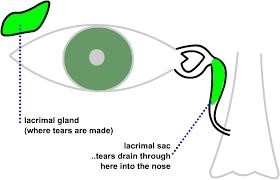
- The Main Lacrimal gland:- It is situated in the fossa of the lacrimal gland in the orbital plate of the frontal bone. The main lacrimal gland is divided by lateral horn of aponeurosis into Superior Orbital Part and Inferior Palpebral Part.
Superior Orbital Part- It is about the size and shape of an almond. It has two extremities( lateral and medial),two borders(anterior and posterior) and two surface(superior and inferior).
Inferior Palpebral Part- It consist of two or three lobules and the size is small.
- Accessory Lacrimal Gland:- It includes
- Glands of Krause
- Glands of Wolfring
- Glands of Caruncle and Plica Semilunaris
- Infraorbital glands
Lacrimal Drainage System:- The lacrimal punctum lies on a small fibrous mound, called the ‘ lacrimal papilla’. Diametre of its opening is 0.2-0.3 mm and directs somewhat posteriorly towards the lacrimal lake. The lacrimal canaliculus is divided into the vertical and horizontal portions. The length of the vertical portion is 2mm and that of the horizontal part is 10mm. More than 95% of the upper and lower canaliculi join to become the common canaliculus to reach the common internal ostium. The lacrimal sac lies within a bony fossa bordered by the anterior and posterior lacrimal crests. When operating in the area of the lacrimal sac, the surgeon may encounter the angular artery and vein which lie 7-8mm medial to the medial canthal angle. If these vessels are cut during dacryocystorhinostomy they may cause troublesome bleeding. In adults the Naso-lacrimal duct measures 12-18mm in length.

Blood Supply of Lacrimal Passage:-
- Arterial Supply:-
- Superior and inferior palpebral arteries.
- Angular artery.
- Infraorbital artery.
- Nasal branches of sphenopalatine artery.
- Venous Drainage :-
- From above- Angular vein and Infraorbital vein.
- From below- Nasal vein.
Nerve Supply of Lacrimal Passage:-
- Lacrimal Sac:- Sensory Nerve.
- Nasolacrimal Duct:-Infratrochlear nerve and Anterior Superior Alveolar nerves.
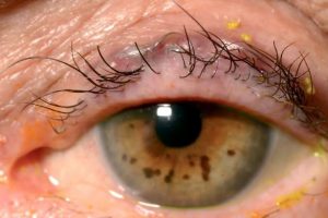
Causes of a watering eye:
Epiphora is defined as the sign of overflow of tears, and may be caused by the
following:
- Hypersecretion (pseudoepiphora) secondary to ocular inflammation or
surface disease. In these cases watering is associated with symptoms of the
underlying cause and treatment is usually medical.
- Defective drainage (true epiphora) due to compromise of the lacrimal
drainage system. This tends to be exacerbated by a cold and windy
atmosphere, and is least evident in a warm dry room. It may be caused by:
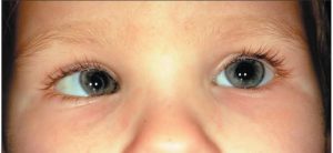
- Malposition of the lacrimal puncta (e.g. secondary to ectropion).
- Obstruction along the lacrimal drainage system, from the puncta to
the nasolacrimal duct
- Lacrimal pump failure, which may occur secondarily to lower lid
laxity or weakness of the orbicularis muscle (e.g. facial nerve palsy).
Pseudoepiphora :- Some patients feel like their eyes have too many tears but do not exhibit frank epiphora. These sensations are often caused by other ocular or eyelid abnormalities. For example, patients with dry eye may perceive foreign-body sensation or increased mucus production as excess tearing, but they do not exhibit true overflow of tears over the eyelid margin or down the cheek.
Lacrimal outflow examination:- Nasal examination may uncover an unsuspected cause of the epiphora, such as an intranasal tumor, turbinate impaction, or chronic allergic rhinitis. These conditions may occlude the nasal end of the NLD.
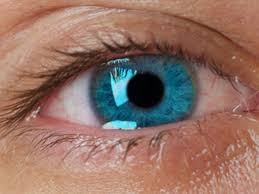
Diagnostic tests:- The dye disappearance test (DDT) is useful for assessing the presence or absence of adequate lacrimal outflow, especially in unilateral cases. It is more heavily relied upon for children, in whom lacrimal irrigation is impossible without deep sedation. Using a drop of sterile 2% fluorescein solution or a moistened fluorescein strip, the examiner instills fluorescein into the conjunctival fornices of each eye and then observes the tear film with the cobalt blue filter of the slit lamp. Persistence of significant dye and, particularly, asymmetric clearance of the dye from the tear meniscus over a 5-minute period indicate an obstruction. If the DDT result is normal, severe lacrimal drainage dysfunction is highly unlikely.


