
Tear film Dynamics is a complex process controlled by the various factors, main and accessory lacrimal glands, ocular surface structure (cornea, conjunctiva ,eyelids with meibomian gland) and interconnecting nerves (both sensory and motor).
🔸Wolff was the first to describe the detailed structure of the fluid covering the cornea and called it Pre corneal film.
Structure
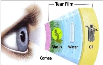
- Wolff described in detail the structure Of the tear film. Tear film consists of three layers which from anterior to posterior are
- Lipid layer/ Oily layer
- Intermediate Aqueous layer
- Mucus layer/ Inner mucin layer.
Mucus layer – It is the innermost layer. 0.2micrometer thick of the tear film ,it can be demo in living eye by alcian blue drops. It is secreted by conjunctival goblet cells and glands of Manz.
Function – It converts the hydrophobic corneal surface into hydrophilic one. It plays a vital role in the stability of the tear film. Absorbs various organic compounds in tears. Lubricates the ocular and palpebral surfaces.
Aqueous layer – It is the middle layer of tear film. Thickness over the cornea 7.0micrometer. comprise of water and small quantities of solutes such as sodium chloride, sugar, urea and proteins. Therefore it is alkaline and salty in taste. Formed by secretions from the main and accessory lacrimal glands of Krause and Wolfring.
Function – Wash away debris and allow the passage of leucocyte after injury. Supply atmospheric oxygen to the avascular corneal epithelium. Contains antibacterial substances like lysozyme, lactoferrin and beta- lysin.
Lipid/ Oily layer – It is the outermost and thinnest layer. 0.1micrometer thick. Derived from secretion of Meibomian, Zeiss and Moll glands. It Contains
- low polarity lipids Like wax
- cholesterol
- esters
- high polarity lipids like TG, FFA, phospholipids
Function – Prevents the overflow of tears. Lubricates the eyelids, prevents evaporation.
Physical properties of tear film –
Thickness – 4-8micrometer, Volume- 7 micrometre( highest bin youth and then gradually decreases), Rate of secretion – 1.2 micrometre per min, PH – 7.3- 7.7
Secretion of tears
Tears are continuously secreted throughout the day by accessory (basal secretion) and main (reflex secretion)lacrimal glands.
Basal secretion – In human eyes the cornea is continually kept moist and nourished by basal tears. They lubricates the eye and help to keep it clear of dust. Secreted by accessory lacrimal glands.
Reflex secretion– Secreted by main lacrimal gland. Results from irritation of the eye by foreign particles. Can also occur with bright light and hot and peppery stimuli to the tounge and mouth.
Formation of pre-ocular tear film
- Wettability of a surface – Conjunctival mucus spreads on to the cornea by the action of the kids. On this new surface aqueous layer spread spontaneously. Then, superficial lipid layer spreads contributing to its stability band regarding evaporation between blinks.
- Retention and Redistribution of tear film – The outermost layer of corneal epithelium along with mucopolysaccharides play an important role in retaining fluid on corneal surface. Precorneal film is stagnant until it blinks or eye movement. Redistribution occurs in the form of bringing of new tear fluid by way of marginal strip where there is constant flow of tears.
- Displacement phenomenon – Cornea’s surface is covered by a film which has stability, compressibility, elasticity and unaffected by gravity. Displacement phenomenon is possible due to thin monocular layer on the surface of cornea.
- Evaporation of the tear film – All lipid fluids including wax esters and cholesterol, esters retard evaporation of water. Evaporation is about 10% production rate, which accounts to about 0.12 microliter/ min.
- Drying and break up of tear film – Pre corneal tear film has a short lived stability. It is re-established completely after a blink. It takes 15-40 sec for tear film to rupture and dry spots to appear. Drying of corneal surface can’t be a result of evaporation of water alone as it takes at least 10mins.
- Elimination of tears – Drainage of lacrimal fluid from Lacus-lacrimalis into Naso-lacrimal duct. Lacrimal fluid flow downwards and laterally across the evaporation from ocular surface . Variable amount of tears is lost by evaporation from ocular surface and remainder of tears flow along the superior and inferior marginal strips and collects as lacus lacrimalis in the inner canthus .Fromwhere, it is drained by the lacrimal passage into the nasal cavity. 70% of tears is drained via inferior canaliculus and 30% via the superior canaliculus by an active pump mechanism.
Lacrimal pump mechanism – Operates with the blinking movements. Performed by orbicularis muscle of eyelid. Two major events –
- eyelid closure
- eyelid open
When eyelid close – Contraction of pretarsal fibres of orbicularis compress the ampulla and shortens the canaliculi. This movement propels the tear fluid present in the ampulla and horizontal part of canaliculi toward the Lacrimal sac. At the same time inferior portion closes more tightly this preventing aspiration of air from nose.
When eyelid open – Relaxation of pretarsal orbicularis muscle allow’s the canaliculi and ampulla to expand and reopen, and to draw the tear fluid from the lacus lacrimalis and marginal tear strips. Relaxation of presepatal fibres results in collapse of sac as a consequence a positive pressure is created which forces the tears down to the naso lacrimal duct into the nose. At the same time puncta moves laterally , canaliculi lengthens and is filled with tears.
- Drainage of lacrimal fluid from NLD into Nasal cavity.
- Gravity helps downwards flow.
- Air currents induce negative pressure in NLD.
- Opens the Hasner’s Valve .
- Draws the fluid down into the lumen of the nose.


