
Conjunctiva is a thin transparent mucous membrane, which lines the posterior surface of the eyelids and anterior surface of eyeball. Conjunctiva is composed of non-keratinized , stratified squamous epithelium with goblet cells , stratified columnar epithelium and stratified cuboidal epithelium. Conjunctiva means ‘conjoin to join’, the name has been given this mucous membrane that joins the eyeball to the lids. It stretches from the lid margin to the limbus , and encloses a complex space called conjunctival sac. A healthy conjunctiva is necessary for the eye to function normally , as it helps to create a suitable environment for the cornea, which is responsible for focusing most of the light that enters the eye. It helps protect the eye by keeping out foreign body and microorganisms and it also maintain tear film. Primary function of the conjunctiva is to keep the front surface of the eye moist and lubricated, and it also keeps the inner surface of the eyelids moist and lubricated that making the eye able to open and close the eye without any irritation. Another function of the conjunctiva is to protect the eyes from dust, debris, pollens and micro organisms that could cause infection. Because conjunctiva contains many small blood vessels , it is able to provide important nutrients to the eye and eyelids. It also contains special cells that work with the tear film that prevent dry eye .
Conjunctiva can be divided into 3 parts
Palpebral Conjunctiva :- This part of conjunctiva is a clear membrane that coats inside the upper and lower eyelids. It can be subdivided into 3 parts .
- marginal conjunctiva ;- This type of conjunctiva extends from the lid margin to about 2 mm on the back of lid upto a shallow grove, this is called sulcus subtarsalis. It is actually a transitional between the skin and conjunctiva proper.
- Tarsal conjunctiva ;- Tarsal conjunctiva is thin, transparent membrane. It is adherent to the whole tarsal plate in the upper eyelid, in the lower eyelid it is adherent only to half width of the tarsus.
- Orbital part of conjunctiva ;– It lies between the tarsal plate and fornix. It lies over muller’s muscle in upper eyelid.
Bulbar Conjunctiva :- Bulbar conjunctiva is a thin, transparent conjunctiva which covers the anterior part of the sclera and it does not cover the cornea. It is separated from the anterior sclera by episcleral tissue and Tenon’s capsule. 3 mm ridge in bulbar conjunctiva around the cornea is called ‘limbal Conjunctiva’. The average thickness of bulbar conjunctiva membrane is 33 microns.
Fornix :- The fornix of conjunctiva is a thin , loose and soft tissue which lying at the junction between the palpebral conjunctiva and bulbar conjunctiva. It can be subdivided into 4 parts
- Superior fornix lies between the upper lid and globe. It extends 8 to 10 mm from the upper border of the limbus.
- Inferior fornix lies between the lower lid and the globe. It extends up to a distance of 8 mm below the part of the limbus.
- Medial fornix is the shallowest and contains the caruncle and the plica semilunaris.
- Lateral fornix lies between the lateral canthus and globe . It extends for a distance of 15 mm from the lateral part of the limbus.
Histology :-
Histologically , conjunctiva consists of three layers
- Epithelium :- Epithelium is a 2-5 layered , non- keratinized epithelium. It also contains goblet cells . Marginal conjunctiva has 5 layered stratified squamous type of epithelium, tarsal conjunctiva has 2 layered of epithelium, fornix and bulbar conjunctiva have 3 layered of epithelium and limbal conjunctiva has many layered of epithelium.
- Adenoid layer :– Adenoid layer conjunctiva is also called lymphoid layer . It consists of fine connecting tissue reticulum in the meshes of which lie lymphocytes. This layer not present since birth ,it develops 3-4 months of life.
- Fibrous layer :– Fibrous layer consists of a meshwork of collagenous and elastic fibres. This layer contains vessels and nerves of conjunctiva.
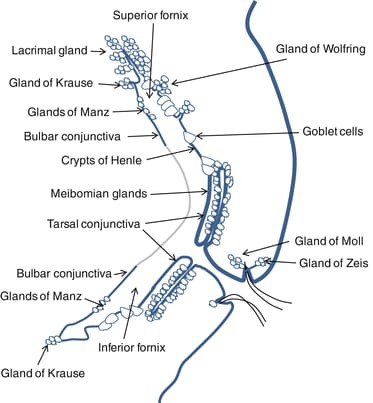
Glands of conjunctiva :-
Two types of glands –
- Mucin secretory glands – These include Goblet cells , crypts of henle and Glands of Manz
- Accessory lacrimal glands – These include Glands of Wolfring and Glands of Krause.
Blood Supply :-
The blood supply of the conjunctiva is from-
The marginal arcade of the eyelid for the marginal conjunctiva
The peripheral arcade of the eyelid for the fornical conjunctiva
The posterior conjunctival artery which is a branch of the peripheral arcade to within 4 mm of the limbus
The anterior conjunctival artery which is a branch of the anterior ciliary for the limbus
The capillary arcades extending 1 mm into the cornea.
Nerve Supply :- The conjunctival nerve supply consists of branches of the ophthalmic division and branches of the maxillary division of the fifth cranial nerve such as superior orbital , infraorbital, supratrochlear , infratrochlear , lacrimal and long ciliary nerve.
Functions of conjunctiva :-
Function of conjunctiva are as follows –
- Tear production
- Supply of oxygen directly to the cornea when the eyes are open.
- Wash off devris.
- Maintain a smooth ocular surface
- Protection of the eye
Microbiology :-
The conjunctival sac is never free organisms, because of its relatively low temperature, evaporation of lacrimal fluid and moderate blood supply, bacteria do not readily propagate themselves. Moreover, the tears are not a good culture medium, and although they contain a bacteriostatic enzyme, lysozyme , they cannot be regarded as actively antimicrobial. Hence they act principally in a mechanical manner, washing away deleterious agents and their products. However, bandaging the eye arrests the movements of the lids and temperature of the sac, thereby increasing the bacterial content of the conjunctival sac.


