
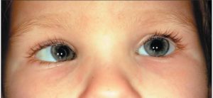
Definition: Normally, visual axis of the two eyes are parallel to each other in the primary position of gaze, and this alignment is maintained, in all position of gaze.
But strabismus is a condition in which only one of the visual axes is directed towards the fixation object, the other being deviated away from this point.
Types:
ESO- Nasal horizontal deviation(relative to fixing eye).
AXO- Temporal horizontal deviation(relative to fixing eye) .
HYPER- Superior vertical deviation (relative to fixing eye) .
HYPO- Inferior vertical deviation(relative to fixing eye).
TROPIA-manifest disorder of ocular alignment.
PHORIA- Latent disorder of ocular alignment.
COMITANT- Ocular deviation only present in specific direction of gaze.
INCOMITANT- Ocular deviation only present in specific directions of gaze
AMBLYOPIA-Clinically defined as a 2-line difference from best corrected visual acuity in a structurally healthy eye.
Classification of squint1) Apparent squint(pseudostrabismus)
2) Latent squint (Haterophoria)
3) Manifest squint (heterotropia)
A) concomitant (non-paralytic)
a) monocular
i) convergent(esoteopia)
ii) divergent(exotropia)
iii) vertical(hypertropia)
Iv) torsional(cyclotropia)
b) alternating
i) convergant
ii) Divergent
B) Incomitant(paralytic)
Symptoms:-
1) Blurring of vision, especially at times of fatigue. Initially, with an effort of fusion, this blurring may be overcome.
2) Poor vision in one eye.
3) Headeche, and eye each are very common.
4) Diplopia or double vision, due to exhaustion of fusional reserve intermittently.
5) There are more white space visible o the outside half of the eyes, the eyes appear to turn inward.
6) one eye always deviated inward and it characteristically develops in early childhood before the binocular reflex are firmly established.
7) one eye always deviated outward. It’s typically seen in older children amd adults.
8) one or both eyes wandering upward or downward..
Primary examinationCommon Examination –
1) Visual acuity testing :- By Snellen’s chart, ‘illiterate E’, Sheridan -Gardiner test, or by special tests, designed for younger children and mentally retards(STYCAR)
2) Refraction under ateopine :- To find out any refractive error.
3) ocular motility :- To find out any limitations of ocular movement.
4) Cover test:-To find out the squint is uniocular or alternating.
5) fixation behavior :- To find out foveal(centric) fixation or eccentric fixation.
6) Anterior segment and fundus :- To rule out any organic lesion which may be the cause of squint.
Examination of a case of heterophoria-
1.Testing for vision and refractive error:-
It is most important, because many times refractive error responsible for the symptoms of the patient or for the deviation itself. So, refraction should be performed under full cycloplegia, especially for childrean.
2.cover-uncover test:-
This is a test that is performed to determine if there is a heterophoria or phoria. To performed it, one eye us covered with an occluder and the other is made to fix an object. In the presence of heterophoria, the eye under covered will deviate. After few seconds later the cover is quickly removed and the movement of the eyeball tels the type of heterophoria. And the speed of movement tells whether recovery is slow or rapid.
Examination of a case of concomitant:-
1.Inspection :- Large degree is obvious or inspection.
2.Ocular Movement :- Uniocular and binocular both movements should be tested in all the cardinal position of gaze.
3.Pupillary Reactions :- These is required because may be abnormal in patients with sensory exotropia due to disease of retina or optic nerve.
4.Testing of vision and refractive error :-
It is most important, because many times refractive error responsible for the symptoms of the patient or for the deviation itself. So, refraction should be performed under full cycloplegia, especially for childrean.
5.Cover test :-There are three types of cover test.
i) Direct cover test :- These confirmed the presence of manifest squint. To perform it, the patient is asked to fixate on a point light. Then the normal looking eye is covered while observing the movement of the uncovered eye. In the presence of squint, the uncovered eye will move in opposite directions to take fixation.
ii) Cover-uncover test :- One eye is covered with an occluder and the other is made to fixate on an object. In the presence of phoria, the eye under cover will deviate. Here, are the eye deviate inwards in cases of exophoria and outwards in cases of esophoria.
iii) Alternate cover test:-It reveals whether the squint is unilateral or alternate (bilateral)
Examination of a case of Incomitant
1.Diplopia Charting :- It’s indicated in patients complaining of confusion or double vision. How it’s perform?
Patient is asked ti wear red and green diplopia charting glasses. Red glass being in front of the right eye and green in front of the left. Then in a semi dark room, he is shown a fine linear light from a distance of 1m, and asked to comment on the images in primary position and in other positions of gaze. Patient tells about the position and the separation of the two images in different fields.
Now note,
i) The direction of gaze, where the separation of images are maximum.
ii) The farther displaced image belongs to which eye. This can be done by covering one eye of the patient, and asking him which one of the two images.
Diplopia chart showing the position of the images in right lateral rectus palsy.
The dotted lines show the position of the false image in different parts of the field of diplopia has disappeared. The farther displaced image belongs to the affected eye.
2.Hess screen (chart) :- The eyes are dissociated similarly with red and green goggles, and note the patient’s response to the dissimilar images formed by each eye while viewing a different targets on Hess Screen.
• The result of the test is interpreted by comparing the grids, charted for each eye and they depend on Herring’s law of equal innervation.
•In paralytic squint, the greatest restrictions occurs in the direction of paretic muscle, with corresponding overaction of the Contralateral synergistic muscle. It also provides the accurate measurement of commitance.
3.Worth’s four-dot test :- Patients eyes are dissociated with red glass placed in front of right eye and green glass in front of left. He then views at a box with four lights, one red 🔴, two green 🟢 and one white ⚪.


