Introduction :
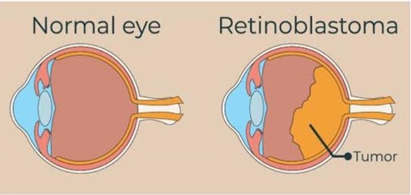
Retinoblastoma is a rare type of eye cancer that usually develops in early childhood, typically before the age of 5 This form of cancer develops in the retina, which is the specialized light-sensitive tissue at the back of the eye that detects light and color. In children with retinoblastoma, the disease often affects only one eye. However, one out of three children with retinoblastoma develops cancer in both eyes. The most common first sign of retinoblastoma is a visible whiteness in the pupil called “cat’s eye reflex” or leukocoria. This unusual whiteness is particularly noticeable in dim light or in photographs taken with a flash. Other signs and symptoms of retinoblastoma include crossed eyes or eyes that do not point in the same direction (strabismus), which can cause squinting; a change in the color of the colored part of the eye (iris); redness, soreness, or swelling of the eyelids; and blindness or poor vision in the affected eye or eyes. Retinoblastoma is often curable when it is diagnosed early. However, if it is not treated promptly, this cancer can spread beyond the eye to other parts of the body. This advanced form of retinoblastoma can be life-threatening. When retinoblastoma is associated with a genetic change (mutation) that occurs in all of the body's cells, it is known as hereditary (or germinal) retinoblastoma. People with this form of retinoblastoma typically develop cancer in both eyes and also have an increased risk of developing several other cancers outside the eye. Specifically, they are more likely to develop a cancer of the pineal gland in the brain (pineoblastoma), a type of bone cancer known as osteosarcoma, cancers of soft tissues (such as muscle) called soft tissue sarcomas, and an aggressive form of skin cancer called melanoma.
Signs & Symptoms :
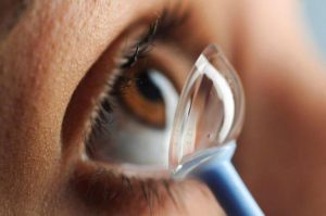
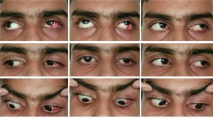
Retinoblastoma may be discovered during a routine exam by a pediatrician; however, most often the parent is the first one to notice signs of retinoblastoma. For the majority of children with retinoblastoma, the sign that is noticed is a white pupil reflex or leukocoria. Leukocoria causes the pupil of the eye to reflect white, as pictured, instead of the normal black (or normal red reflection in a flash photograph).Other eye diseases can also cause this white pupil reflex, so leukocoria does not always indicate retinoblastoma. An ophthalmologist can determine the correct diagnosis. A crossed eye or strabismus is the second most common sign of retinoblastoma. The child’s eye may turn outward (towards the ear) or inward (towards the nose).Retinoblastoma may also be noticed because of a red painful eye, poor vision, inflammation of the tissue around the eye, an enlarged (dilated) pupil, or a different colored iris. Retinoblastoma may
cause other symptoms, like a sudden decrease in eating or drinking.
Classification :
The severity of retinoblastoma tumors may be classified by either of two systems: the Reese-Ellsworth classification system and the International Classification. The higher the Group number or letter in the system, the poorer the prognosis is for saving the eye. Some centers may use one classification over the other, but at MSK we use both.
Reese-Ellsworth Classification for Retinoblastoma:
This classification system was developed as a method to predict whether the child’s eye can be saved.
GROUP I –
A. Solitary tumor, less that 4 disc diameters in size, at or behind the equator.
B. Multiple size tumors, none over 4 disc diameters in size, all at or behind the equator.
GROUP II –
A. Solitary tumor, 4 to 10 disc diameters in size, at or behind the equator
B. Multiple size tumors, 4 to 10 disc diameters in size, all at or behind the equator
GROUP III-
A. Any lesion anterior to the equator.
B. Solitary tumors larger that 10 disc diameters behind the equator.
GROUP IV-
A. Multiple tumors, some larger than 10 disc diameters
B. Any lesion extending anteriorly to the ora serrata
GROUP V-
A. Massive tumors involving over half of the retina
B. Vitreous seeding. International Classification-
GROUP A-
• Small tumors (less than 3 mm) that are only in the retina and more than 3 mm away from the foveola (the center of the fovea) and more than 1.5 mm away from the optic disk.
GROUP B-
• Tumors larger than 3 mm that are confined to the retina in any location.
• Clear subretinal fluid less than 6 mm from the edge of the tumor.
GROUP C-
• Localized vitreous and/or subretinal seeding (less than 6 mm from the tumor margin).
• No tumor masses, clumps or snowballs in vitreous or in the subretinal space.
GROUP D-
• Diffuse vitreous and/or subretinal seeding (more than 6 mm from tumor).
• Subretinal fluid more than 6 mm from tumor margin.
GROUP E-
• No visual potential OR presence of one or more of the following:
• Tumor in the anterior segment
• Tumor in or on the ciliary body
• Neovascular glaucoma
• Vitreous hemorrhage obscuring the tumor or significant hyphema
• Phthisical or pre-phthisical eye
• Orbital cellulitis-like presentation
Causes :
Mutations in the RB1 gene are responsible for most cases of retinoblastoma. RB1 is a tumor suppressor gene, which means that it normally regulates cell growth and stops cells from dividing too rapidly or in an uncontrolled way. Most mutations in the RB1 gene prevent it from making any functional protein, so cells are unable to regulate cell division effectively. As a result, certain cells in the retina can divide uncontrollably to form a cancerous tumor. Some studies suggest that additional genetic changes can influence the development of retinoblastoma; these changes may help explain variations in the development and growth of retinoblastoma and other types of tumors in different people.
A small percentage of retinoblastomas are caused by deletions in the region of
chromosome that contains the RB1 gene. Because these chromosomal changes
involve several genes in addition to RB1, affected children usually also have intellectual disability, slow growth, and distinctive facial features (such as prominent eyebrows, a short nose with a broad nasal bridge, and ear abnormalities).


