
Wolff was the first to describe the structure of the fluid covering the cornea and called it as precorneal film. He described that the tear fim consist of three layers,which from posterior to anterior are Mucus Layer,Aqueous Layer & Lipid or Oily Layer.
Tear Layer is an important aspect for estimating the suitability of the contact lens wear.
Tear Film Dysfunction or Tear Film Adequacy is a disturbance in the tear film function owing to change in lipid layer,water or mucin components of the tears. Test for tear fim adequacy include –
SPECIAL CLINICAL TESTS :
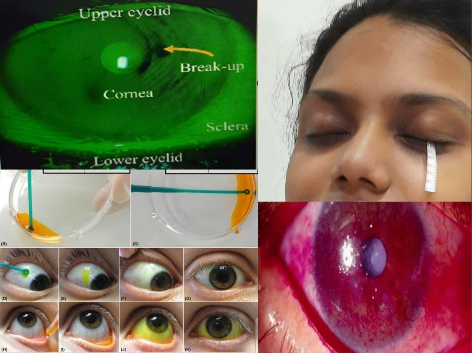
- Tear-Flim Break-Up Time
Tear-Fim Break-Up Time is simple test to assess the stability of the pre-corneal tear fim. A normal TBUT is more than 10 seconds (11-35 seconds), and a TBUT value less than 10 seconds is abnormal.
Procedure for TBUT :
- Drop of fluorescein is instilled and the patient is asked to blink 2-3 times to distribute the dye.
- The patient is then asked not to blink while the cornea is studied by the cobalt blue filter with the slit lamp.
- The TBUT is the time in seconds between the last blink and the appearance of dry(black) spots on the cornea.
- Schirmer’s Test
Schirmer’s test measures the volume of the tears . There are types of schirmer’s test used to measure the volume of the tears are –
- Schirmer’s Test 1 : This test is done without anesthesia. It measures the total secretion( i.e.,basic + reflex).
Schirmer’s test 1 may also be done after topical anesthesia,which measures only the basic secretion.
Values of Schirmer’s Test 1
NORMAL VALUE : 11-25mm
BODERLINE : 5-10mm
IMPAIRED SECRETION : LESS THAN 5mm
Procedure of Schirmer’s Test 1
- 5 mm wide and 35 mm long special filter paper ( Whatman no.41) is placed in lower fornix at the junction of the middle and outer- third of the eyelid after folding it at 5mm.
- After 5 minutes,the amount of wetting from the fold is measured.
- Patient may blink or close the eyes as necessary during the test,
- Schirmer’s Test 11 : This test is done with topical anesthesia. It measures the reflex secretionThis test is seldom used.
Procedure of Schirmer’s Test 11
- The procedure is same except, instillation of topical anesthesia,in the eye and irritation of ipsilateral nasal mucosa with a camel hair brush or cotton swab during the test.
- The reading is taken after 2minutes and a measurement of less than 15 mm indicates failure of reflex secretion .
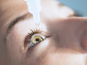
- Vital Dye Staining Test
- Fluorescein (2%) Staining : Fluorescein stains precorneal tear-film and intercellular tissue. It doesn’t stain the mucus and devitalized epithelial cells.
- Rose Bengal (1%) Staining : Rose Bengal staining has an affinity for devitalized epithelial cells, mucus and filaments. It is very useful to detect mild cases of dry eye by staining the interpalpebral conjunctiva in the form of two triangles with their bases towards the limbus. Topical anesthesia should not be used prior to Rose -Bengal staining, as it may induce false-positive result.
- Lissamine Green : Lissamine green staining is used to stain the surface similar to Rose – Bengal stain.
LABORATORY TEST :
- Tear Lysozyme Assay : The test is estimated by placing a tear-wetted filter strip in an agar plate containing the bacteria Micrococcus Lysodeikticus,and zone of lysis is measured after incubation at +37 degree celcius for 24 hours. In KCs there is reduction in tear lysozyme.
- Tear Osmolarity : Normal range is 290-311 mOsm/L,and it increases in KCs. Tear osmolarity value above 316 mOsm/L is diagnostic of dry eye disease.
- Fluorescein Dilution Test : One drop of Fluorescein is instilled in the eye and the dilution over a period of time is measured photometrically to calculate the tear volume.
- Tear Mucin Measurement: Measuring the Hexosamine content (one of the principles moieties of mucus) of tears.
- Globet -Cell Count of the Conjunctiva: Biopsy specimen is taken from the inferior nasal conjunctival fornix for globet cell count test .Average globet -cell count in normal eyes is 8-15 cells/sqmm.The number is diminished in mucin deficiency states.


