
To see an object clearly we need a normal eye alignment that would help to focus light on the macula binocularly. If there remain any irregularities in eye alignment that would create ‘ abnormal retinal correspondence ‘ and further it creates amblyopia in children and diplopia in adults.
ANATOMY OF NORMAL EYE ALIGNMENT –
Eye rest on a facial hammock ( tendinous sheath ) frontally and adipose tissue of the orbit and its movements r controlled by 6 extraocular muscles(EOM) .
1. 4 rectus – superior,inferior,lateral,medial
2. 2 oblique – superior ,inferior

Origin – 4 recti from common ring tendon .superior oblique from sphenoid bone and inferior oblique from maxilla bone .
Insertion – 4 rectus at in front of the equator or near the limbus ( medial rectus ,inferior rectus ,lateral rectus, and superior rectus accordingly 5.5 ,5.9,6.5 and 7.7 mm behind the limbus ) . 2 obliques behind the equator.
Rectus comes from the word recti that means a straight line .But that is not exactly square with the optical axis rather than it makes some angle with it .

Superior and inferior rectus makes an angle of 23° with an optical axis . Medial and lateral rectus r parallel to optical axis . And the two oblique muscles make an angle of 51° with optical axis .
Greater the angle =>Geater deviation from 1° function.
Oblique muscles got greatest angles so they got 1°,2°,3° of equal severity.
Superior and inferior Rectus got mid range angles so they got severe 1° ,mild 2° and low intensity 3° functions
Lateral and medial rectus r parallel to optical axis so they only got 1° function .


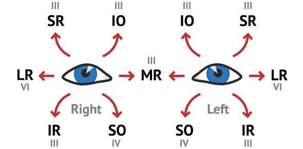
Eye Muscles with their actions –

Types of eye movements –
1. Duction or Uniocular eye movement – These are of 6 types . Adduction or inward movement , abduction or outward movement, supraduction or upward movement , infraduction or downward movement , incycloduction or intorsion ( 12’o clock position rotates towards 3’O clock position ) and excycloduction (12’O clock position moves towards 9’O clock position )
2. Binocular movements – These are of two types 1. Version ,where two eyes moves symultinously in same direction ,and 2. Vergence – where two eyes moves symultinously in opposite directions .
Version are of 10 types – dextroversion or right gaze , levoversion or left gaze , supraversion or upward gaze , infraversion or downward gaze , dextroelevetion right and up gaze , dextrodepression or right and down gaze , levoelevation or left and up gaze , levodepression or left and down gaze , dextrocycloversion or right gaze where 12’O clock position moves towards 3 ‘O clock position , levocycloversion or just opposite of dextrocycloversion .
Vergence are of 2 types – Convergence or simultaneous inward movement of eyeball , divergence or symultinous outward movement of eye ball .
Physiology of ocular movements
Nerve supply of extraocular muscles –
All 3 LR 6 SO 4
That means All the extraocular muscles are supplied by occulomotor nerve except Lateral rectus which is supplied by abducent nerve and Superior oblique which is supplied by trochlear nerve .
Kingdom of occulomotor –

Kingdom of abducent –

Kingdom of trochlear –

Diagnostic positions of gaze –
1.primary – straight vision
2. Secondary – straight up down and right ,left
3. Tertiary – dextro elevation and depression , levo elevation and depression.
4. Cardinal – ‘H’ shaped movements of eyeball

Agonist , antagonist , yoke muscles and contralateral antagonist –
1. Agonist and synergist means …two muscle that helps to move our eye in same direction . This term is used for Uniocular system . eg inferior oblique and superior rectus of same eye .
2. Antagonists means Uniocularly they acts opposite to each other .eg medial and lateral rectus of same eye
3. Yoke muscle or contralateral synergists means one muscle from each eye ,when the acts together they causes version or same direction movement of eye .eg right LR and left MR
4. Contralateral antagonist – pair of muscles ( one from of each eye ) that acts oppositey . eg right LR and left LR .
Laws of ocular movements –
1. Herings law of equal innervation – yoke muscle receives equal and symultaneous innervation from the brain during different binocular movements. eg during convergence both medial recti get equal innervation .

2. Sherringtons law of reciprocal innervation – increased flow of innervation to the contracting muscle is accompanied by decreased innervation to the relaxing antagonist muscle .
Supranuclear control of eye movements
We move our head regualry , in any direction ,with our will. But we see images without any defect .Our brain plays an important role to clearify images despite of regular and irregular head movements and body movements. This is called supranuclear control . This mainly includes – 1. Saccade 2. Smooth pursuit 3. Vestibular system
1.Saccade – when images pass out rapidly our ocular system adjust itself by rapid voluntary movement of our eye . This is controlled by connection between abducent nerve and posterior partial lobe . Eg , viewing object from a fast going train
2. Smooth persuit – when we see a flying bird in the sky our eye moves slowly by following it . This type of slow , following movement of eye is called smooth persuit movement . This is controlled by PPRF ( posterior pontine reticulate fibre ) and persuit control part of frontal brain . Recent study also supports that superior colliculus also supports smooth persuit movement.
Vestibulocular system – Movement of eye with response to gravity and movement is called vestibulocular movement . Semicircular canal , saccule and utricle of inner eye detects the movement and send it’s detection by a complex pathway made by vestibular nucleus ( pontine ) , occulomotor nucleus and abducent nucleus . This prevent retinal image slip during head and body movements .
Movement of eye and its control by neural system is a complicated study and it’s further research is going on . Any recent invention will be notified to you . Keep eye on our page .


