
The extraocular Muscle or extrinsic eye muscle are striated and distinguished from the intrinsic eye muscle which are non-striated and responsible for controlling the movement of iris (dillator and sphicnter pupillae). The fibres of the main muscle mass are also termed extrafusal to distinguishing them from the intrafusal fibres of the central oculomotor regulatory centres. A set of six extraocular muscle (4 recti and 2 obliques) controls the movement of each eyes. Four muscles that run almost a straight course from origin to insertion are called recti muscles and two muscles that a diagonal course are called oblique muscle.
Actions of Extraocular Muscle
There are four types of ocular movement (action)
i) Ductions
ii) Versions
iii) Vergences
iv) Supranuclear movement
Ductions: it refers to the movement of one eye and consist of
a) Adduction(eye movement towards nose)
b) Abduction (eye movement away from nose)
c) elevation(up ward movement of eye)
d) depression( downward movement of eye)
e) intorsion( nasal rotation of eye)
f) extorsion(temporal rotation of the eye)
Versions: these are binocular movement in which the two eyes move synchronously and symmetrically in the same direction.
These include
a)dextroversion (right gaze)
b)levoversion (left gaze)
c)supraversion (up gaze)
d)infraversion (down gaze)
e)dextroelevation ( gaze up and right)
f)dextrodepression (gaze down and right)
g)levoelevation (gaze up and left)
h)levodepression (gaze down and left)
i)dextrocycloversion (rotation to left)
j)levocycloversion ( rotation to right)
Vergences:They are binocular movement in which the two eyes move synchronously and symmetrically in the opposite directions.
Supranuclear movements: the Supranuclear control of the eye movement are the basic ocular movement which must function simultaneously. They are as follows
a) Saccades: they are the rapid eye movement which generally voluntary refixation movement. i.e. to move both eyes quickly from one object to another
b) Pursuits: they are smooth following movements which maintains the visual axes on any slow moving objects.
c) Vestibuloocular movement : they are the co-ordinated eye movement with respect to the gravity and head position.
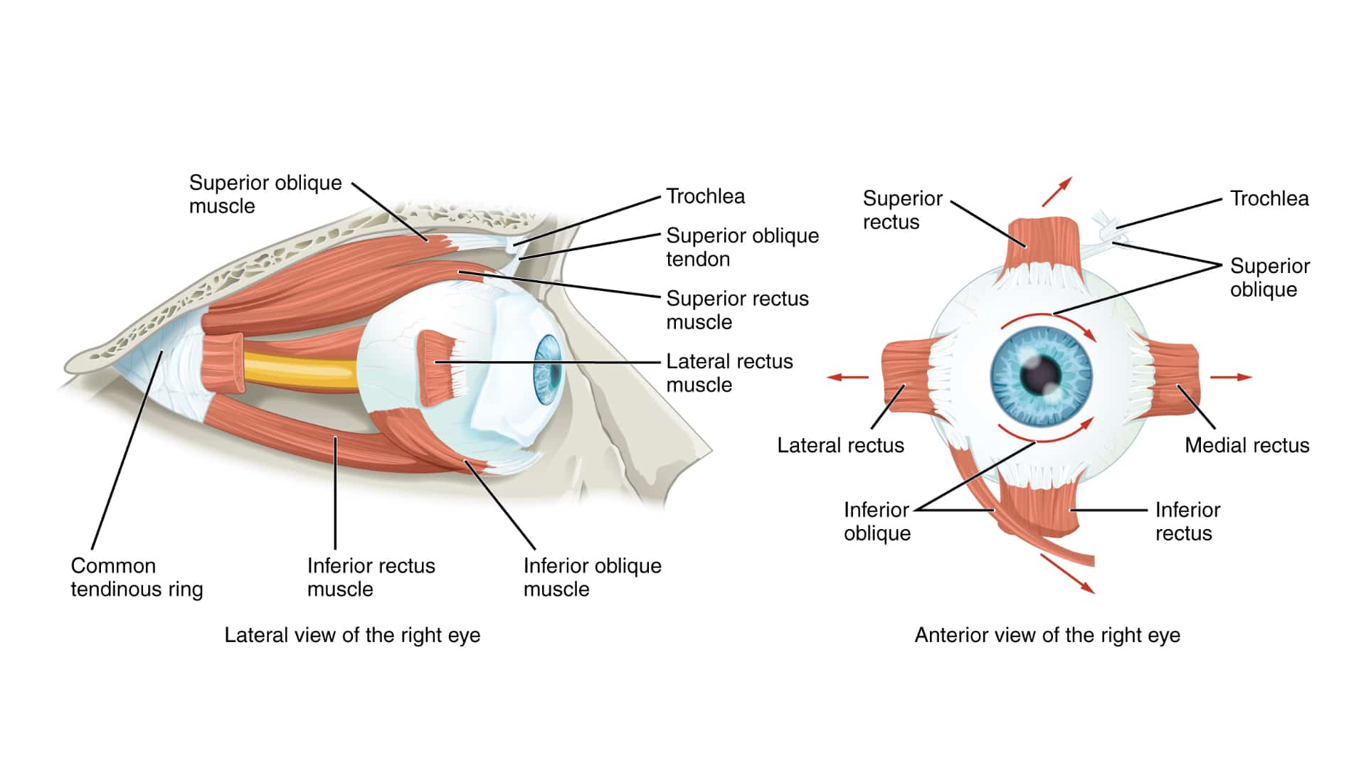
The Recti Muscles
The four recti muscles, superior,inferior,medial and lateral are attached by a fibrous ring of connective tissue located posteriorly by a short tendinous ring(annulus of Zinn).
Superior Rectus: it arises from the upper part of the tendinous ring, pierces Tenon’s capsule and is attached to sclera 7.7mm from the cornea(limbus) by a tendon 5.8mm long and width 10.8mm. This muscle is about 42mmin length and 9mm in width.
Blood supply: the lateral mascular branch of the ophthalmic artery.
Nerve Supply: oculomotor nerve (CN III)
Primary Action: Elevation
Secondary Action: Intorsion
Tertiary Action: Adduction
Inferior Rectus: it is the shortest of the recti and is attached to the inferior aspect of tendinous ring. It is attached to the sclera 6.5mm from the cornea(limbus) by a tendon 5.5mm long and width 9.8mm. This muscle is about 40mm long and 9mm wide.
Blood Supply: the medial muscular branch of the ophthalmic artery
Nerve Supply: oculomotor nerve (CN III)
Primary Action: Depression
Secondary Action: Extorsion
Tertiary Action: Adduction
Medial Rectus: it is the largest Ocular muscle and stronger than the lateral rectus. It is widely attached medial and inferior to the tendinous ring by both part of the common tendon and the sheath of the optic nerve. It is 40mm long and thicker than the any other extraocular muscle. It passes along the medial wall of the orbit and inserts into sclera 5.5mm from the cornea(limbus) by a tendon 3.7mm in length.
Nerve Supply: oculomotor nerve (CN III)
Blood Supply: the medial muscular branch of the ophthalmic artery.
Action: Adduction
Lateral Rectus: it arises from the lateral aspect of the tendinous ring. It is about 48mm in length which is longer than medial rectus but only two-thirds of its cross-sectional area. It reaches sclera 6.9mm from cornea(limbus) by a tendon 8.8mm long. The lateral rectus is often visible through the conjuntiva and Tenon’s capsule.
Nerve Supply: Abducens nerve (CN VI)
Blood Supply: the blood is supplied by the lacrimal artery and lateral muscular branch of the ophthalmic artery.
Action: Abduction
Oblique Muscles
There are two oblique muscles, the superior and inferior oblique. They originate from the bones of the orbital cavity.
Superior Oblique: it arises from the bone (body of sphenoid). It is the longest and thinnest extraocular muscle.
Nerve Supply: trochlear nerve (CN IV)
Blood Supply:it is served by the superior muscular branch of the ophthalmic artery.
Primary Action: Intorsion
Secondary Action: Depression
Tertiary Action:Abduction
Inferior Oblique: it is the only extraocular muscle attached near the front of the orbit and has the tendon of insertion. Some of its muscle fibres are directly attached to sclera.
Nerve Supply: oculomotor nerve (CN III)
Blood Supply: it served by the infraorbital and medial muscular branch of the ophthalmic artery.
Primary Action: Extorsion
Secondary Action: Elevation
Tertiary Action: Abduction


