
The eyeball is a bilateral and spherical organ , which house the structures responsible for vision . It lies in a bony orbit . Anatomically, the eyeball can be divided into three parts – the fibrous , vascular and inner layers . There are six extraocular muscles ( four rectus and two oblique ) . Origin of the rectus muscle from annular tendon of zinn around the optic foramen at the apex of the orbit . Origin of superior oblique from annular tendon of zinn then runs to the trochlea at upper and inner angle of orbit then reflected backward under the superior rectus muscle . Origin of the inferior oblique anteriorly from the lower and inner orbital walls near the lacrimal fossa . It is the only muscle which doesn’t arrived from the lower from the tendon of zinn . These extraocular muscles are responsible for movements of the eye along three different axis : horizontal , either toward the nose ( adduction ) or away from the nose ( abduction ) ;and torsional movement that bring the top of the eye toward the nose ( intorsion ) or away from the nose ( extorsion ) . Versions are binocular movements in which the two eyes moves synchronously and symmetrically in the same direction . These movements are dextroversion , levoversion , dextroelevation , levoelevation , dextrodepression and levodepression . These are called six cardinal position of the gaze . If there is any problem to move the two eyes in the same direction then problem is detected . These eye movements are possible for yoke muscle . Yoke muscle is a muscle of one eye is paired with a muscle of the opposite eye , while moving the eyes into each of the six cardinal position of the gaze. For example , in dextroversion, two yoke muscles are right lateral rectus and left medial rectus . In dextroelevation , the yoke muscles are right superior rectus and left inferior oblique .
Binocular Single Vision –
Binocular single vision ( BSV ) is the coordinate use of the two eyes , in order to produce a single unified image in three dimensions. There are three types of grade in BSV ; they are – SMP , Fusion , Stereopsis .
- Examination of eyeball
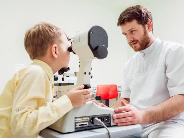
- Synaptophore – is an ophthalmic instrument which is used for diagnosing the imbalance of the eye muscle and treating the by orthoptic methods . It is an instrument which compensates for the angle of squint and allows the stimuli to be presented to both eyes simultaneously .
- It is used to investigate the potential for binocular function in the presence of manifest squint .
- Specially used in children
- Also used to detect suppression and abnormal retinal correspondence
- It is used to assess the angle of deviation and binocular potential at a theoretical distance fixation .
- It is used to test SMP , Fusion , Stereopsis
Work procedure –
- Consist of two cylindrical tubes with a mirrored right angle blend
- A +0.65D lens in each eyepiece which optically sets the testing distance to about 6 meters
- Pictures are interested in a slide carrier situated at the outer end of each table
- Two tubes are supported on columns which enable the pictures to be moved invelation to each other
- It can measures horizontally , vertically and torsional misalignedments simultaneously
- Cover test –
Cover testing is the gold standard for the objective method for determining the presence , type and magnitude of ocular misalignment
▪︎ cover uncover test – The cover-uncover test is generally performed first. The cover-uncover test is useful to identify a tropia and differentiate it from a phoria .
▪︎ Alternate cover test – The alternate cover test is generally performed after the cover-uncover test. The alternate cover test is the most dissociative cover test and measures the total deviation, which includes the tropia plus the phoria .
- Corneal reflection test – A reasonable assessment
of the angle is made by holding a fixation light at 30 cm distance from the patient, and estimating the distance of the corneal light reflex (Purkinje’s first image) from the pupillary center.
One mm decentration of reflex corresponds t o about 7° of ocular deviation. For example, if the reflex is situated at the pupillary margin, the angle is about 15°, and if it is at the limbus, the angle of squint is about 45°. This test is also useful to differentiate it from pseudostrabismus.
- Prism reflex test – prisms are placed in front of the deviating eye until the corneal light reflex become symmetrical
- Prism bar cover test – The prism power required to negate the prism required to negate the ocular movements , equals to the angle of deviation , and it is measured in prism diopter .Roughly, 1° of ocular deviation equals to 2 prism diopter deviation . It is the most accurate method of measuring the angle of deviation .


