
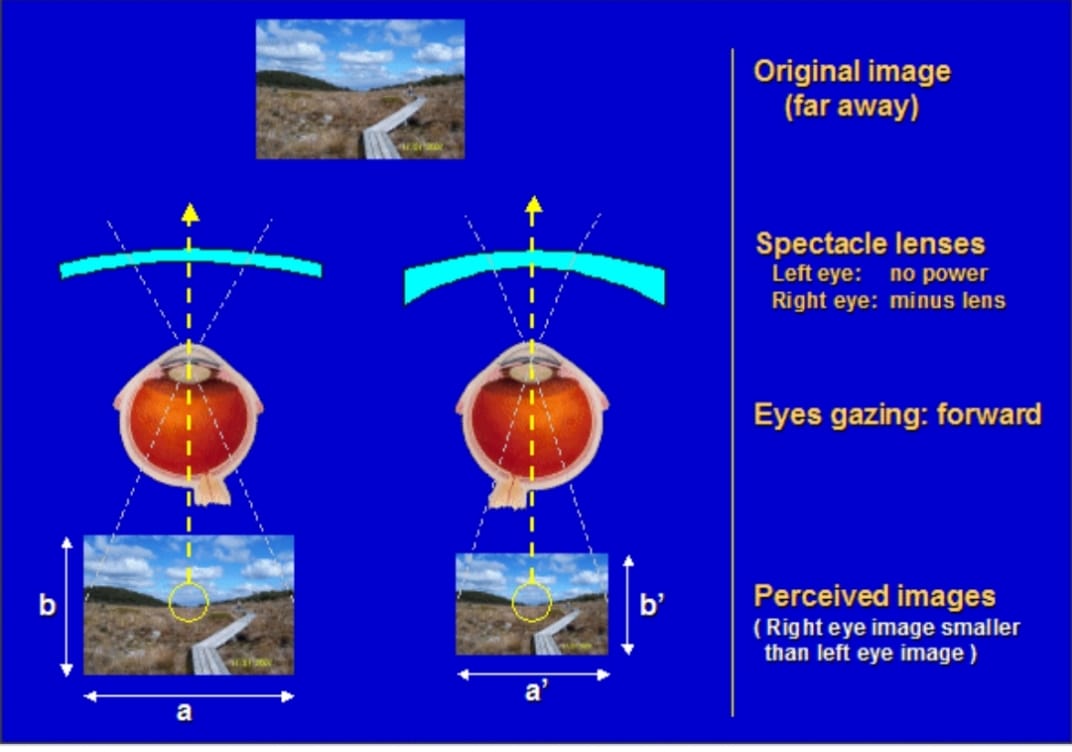
In older optometric/ophthalmic textbooks, rules of thumb have been defined to correct aniseikonia, without actually testing for the amount of aniseikonia. These rules are based on Knapp’s law, which deals with an image size difference as projected onto the retina in anisometropia (i.e., only optical effects are taken into account). Eye care professionals using these rules will base treatment on the patient’s prescription and perhaps the difference in corneal curvature or eye length between the eyes. However, in the more recent literature it has been well established that even in anisometropia, the retinal receptor distribution may also play a role,10-14 and therefore these rules of thumb should not be used. Instead the aniseikonia should be measured.
There are basically two methods to test for aniseikonia: the space eikonometric method and the direct comparison method. The space eikonometric method is based on binocular space perception, while the direct comparison method is based on directly comparing perceived image sizes between the two eyes.
Space Eikonometer:
Methods: Eighteen subjects, ages 21 to 61 years, with normal binocular vision and normal visual acuity had aniseikonia measured with both the Aniseikonia Inspector Version I and the Space Eikonometer. Aniseikonia was measured first with the subjects’ habitual refractive correction and then with afocal size lenses of 1%, 2%, and 3.5% added in random order before the right and left eyes. Measurements were taken initially with the Aniseikonia Inspector and on a subsequent day with the Space Eikonometer.
For the Space Eikonometer, the slopes of the lines for the relationship between the measured aniseikonia and the induced magnification differences in the vertical and horizontal meridians are not significantly different from 1.0. For the Aniseikonia Inspector, the slopes of the lines in the vertical and the horizontal meridians are less than and significantly different from 1.0. On average, the Aniseikonia Inspector underestimates the magnitude of induced aniseikonia, predicting only 68% and 61% of the overall size lens magnification in the vertical and horizontal meridians, respectively. The corresponding values of the Space Eikonometer in the vertical and horizontal meridians are 99% and 93%. Variability is greater with the Space Eikonometer than the Aniseikonia Inspector.
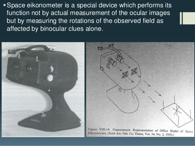
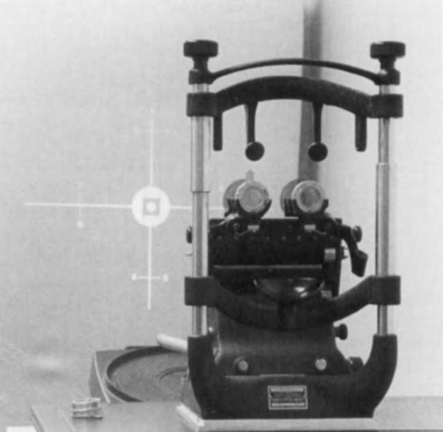
Standard Eikonometer: The standard Eikonometer is a projection instrument that uses polarizing optics to segregate right & left eye views. A focused cross & fixation target is viewed with both eyes. Along the arms of the cross are Nonius lines. If the Nonius lines for the right & left eyes coincide, there is no Aniseikonia.
In the presence of Aniseikonia, the Nonius lines are mismatched along the horizontal or the vertical axes of the cross. This mismatch can be nulled with the use of size lenses. This instrument does not depend on the stereoscopic effect. American Optical space eikonometers are no longer manufactured & today are probably mostly available in educational settings.
Direct Comparison Test:
The New Aniseikonia Test (NAT) is a direct comparison test of perceived image size differences. It uses red/green filter to produce dissociation. The patient compares the size of adjacent right and left images, and differences can be nulled with size lenses. The images viewed by one eye can be altered in size relative to the other.
Others type measurements
Maddox Rod:
One method for measuring Aniseikonia involves the use of a Maddox Rod before one eye. This method was first described by Brecher. As the patient views two penlights. One eye sees streaks of light, while the other sees the penlights. If there is a magnification difference between the eyes, the streaks & the penlights are separated by an amount proportional to the Aniseikonia.
Size lenses are interposed to equalizer the distance, thereby directly estimating the image size difference between the eyes. This procedure could be repeated for different meridians by simply rotating the Maddox rod & the penlights
Red & Green filter: Red & Green filter are used rather than Maddox rod to isolate the two eyes views. Comparisons between the separation between the red and green lights are then the subjective indication of size differences. Size lenses are placed in front of the eye & is kept increasing till image separation is overcome.


