
The human eye is a complex optical system with multiple axes and angles. Varieties of axes and angles of the eye have been defined over the years. Some of the definitions and explanations are practical while others exist only in theory. The anatomical axis is the line passing from the posterior pole to the centre of cornea. Nodal point is where all the light rays go undeviated. The centre of rotation of the eye is the point around which the eye performs rotator movements. Broadly the eye has three principal axes and three visual angles.
- Optical axis
- Visual axis
- Fixation axis
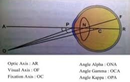
- Optical axis : is the line passing through the centre of the cornea (P) and centre of the lens (N) and meets the retina (R) on the nasal side of the fovea . in practice it is impossible to determine accurately the optical axis, since we cannot know the exact centre of cornea. However, it is much easier to estimate centre of the pupil, for example, by the image of the light on cornea. Therefore, in practice we substitute the optic axis by a line perpendicular to the cornea and the point coinciding to the centre of the pupil. This line is called the pupillary line. The optical axis is only a theoretical concept that applies best to eye models and schematic eyes where the refracrive surfaces centered with respect to one another.
- Visual axis : is the line joining the fixation point (O), nodal point (N) and fovea (F). visual axis is one of the most discussed axes of the eye and is also known as the foveal- fixation axis.
- Fixation axis :is the line joining the fixation point (O) and the centre of rotation of the eye (C).
ANGLES OF THE EYE
1)Angle alpha: is the angle (ONA) formed between the optical axis (AR)and the visual axis(OF) at the nodal point. Normally the visual axis cuts the cornea on nasal side of the optic axis. It is a positive angle alpha (about 5 degrees) and causing an ‘apparent squint’. It may be negative when the visual axis cuts the cornea on the temporal side.
2)Angle gamma :is the angle (OCA) formed between the optical axis (AR) and fixation axis (OC) at the centre of rotation of the eye (C).
3)Angle kappa : is the angle (OPA) formed between the visual axis (OF) and pupillary line (AP). The point P on the centre of cornea is considered equivalent to the centre of pupil.
Practically only angle kappa can be measured (using synaptophore /orbscan) and is of clinical significance. The average angle kappa is 5degree . a positive angle kappa(large) results in pseudo-exotropia and a negative angle kappa in pseudo-esotropia. The measurement of angle kappa is of paramount consideration in refractive surgery, as proper centration is required for optimal results.
CLINICAL IMPLICATIONS OF ANGLES AND AXES OF THE EYE
Hyperopic LASIK refractive surgery :A systemic review of multiple studies reavealed that , in patients with significant hyperopia, centering on the corneal light reflex results in better corrected and uncorrected visual acuity, less decentration of the ablation zone and less higher order aberrations. The same conclusions cannot be made for eyes with mild hyperopias with smaller angle kappa.
Myopic LASIK refractive surgery :according to okamoto et al approximately 1/3 rd of myopic LASIK candidates have an angle kappa large enough to warrant centering closer to the visual axis.
Cataract surgery : studies have shown that eyes with large angle kappa are correlated with more glare and more halos after cataract surgery with placement of multifocal IOLs .
SMILE (small incision lenticule extraction): the adjustment of the intraoperative angle kappa during the SMILE procedure was found to result in few higher-order aberrations.
Macular drag : because fovea is one of the terminal points of visual axis, any pathology that disrupts the foveal anatomical location can lead to alterations to this axis. In pathologies such as choroidal—neovascularisation from ARMD , the contour and location of the fovea are altered. Altered optical axes of the eye are more commonly seen in pathologies causing drag of fovea such as in ROP or Familial exudative vitreo-retinopathy.
References : optics and refraction (AK Khurana)
Essentials of ophthalmology (SK Basak)
Health kura


