

Introduction: The tear film covers the ocular surface and is essential for
protecting the eye from the environment, lubricating the ocular surface,
maintaining a smooth surface for light refraction, and preserving the
health of the conjunctiva and the avascular cornea.
The tear film is approximately 3 to 10 μL in volume, 3 μm thick, and
secreted at a rate of 1 to 2 μL/min. The pH of tears is approximately 7.45
and ranges between 7.14 to 7.82, depending on diurnal and seasonal
influences. Prolonged lid closure, such as during sleep, leads to a buildup
of carbon dioxide, thus lowering the pH. It can conceptually be thought
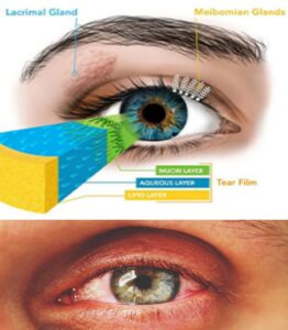
of as having three major layers – inner mucin, middle aqueous, and outer
lipid layer, each of which has separate functions .
The outer lipid layer: It secreted by the meibomian glands, glands of zeis
and glands of Moll.
Function of outer lipid layer:
1. To retard the evaporation of aqueous layer of the tear film.
2. To increase surface tension and vertical stability of the tear film to
prevent overflow of tears.
3. To lubricate the eyelids.
The middle aqueous layer: It is the main bulk of the tear film and secreted
by the lacrimal and accessory lacrimal glands.
Function of middle aqueous layer:
1. To supply atmospheric oxygen to the corneal epithelium.
2. It has antibacterial activity due to presence of lysozyme and
lactoferrin.
3. To provide smooth optical surface.
4. To wash away dust and debris from the conjunctival and corneal.
The inner mucin layer: It is secreted by the conjunctival goblet cells, glands
of Manz and crypts of henle.
Functions of inner mucin layer:
1. To convert the corneal epithelium from a hydrophobic to a hydrophilic
surface.
2. To wet the microvilli of the corneal epithelium and thereby retains the
precorneal tear film.

The main lacrimal glands produce most of the aqueous tear layer, with
small amounts produced by the goblet cells in the conjunctiva and
accessory lacrimal glands.[2] The tears then evaporate or are drained
through the lacrimal puncta.
Another time, there are three different types of tears, each with
unique biochemistries.
Basal tears are typically present on the ocular surface, providing
nutrients to the ocular surface, maintaining ocular comfort, and
ridding the surface of debris.
Reflex tears are those released in response to irritants, including
chemicals and foreign bodies. Reflex tears are produced in
higher quantities than basal tears and are involved in flushing
the ocular surface of irritants.
Closed eye tears are those lubricating the eyes during sleep.
Some components of the tear film, such as lactoferrin, lipocalin-1, and
lysozyme, remain relatively constant between different types of
tears. However, the total amount of protein, lipid, and secretory IgA varies between types; protein and lipid content is highest in basal
tears. Despite differences in composition, the osmolarities in tear types
remain relatively constant.
Functional significance of Tear film
Functions of the tear film include providing lubrication to the ocular
surface and eyelid, antimicrobial defense, providing a smooth ocular
surface for refraction, and supplying oxygen and nutrition to the
avascular corneal epithelium.
The tear film forms a protective barrier between the ocular surface
and the external environment, and it contains antimicrobial
properties. Key antimicrobial factors found in the tear film include
lysozyme, lactoferrin, transferrin, ceruloplasmin, IgA, IgG, IgE,
complement, glycoprotein, and anti-proteinase, which are found in
the aqueous layer of the tear film.
Lysozyme is bacteriolytic, hydrolyzing bacterial peptidoglycan cell
walls. It is highest in concentration in tears compared to other bodily
fluids. Lactoferrin chelates iron, sequestering it from the bacteria that
require it for metabolism and growth. Immunoglobulins play a key role
in defense against bacterial, viral, and parasitic infection. IgA levels are
increased during infectious or inflammatory states, including acute
bacterial conjunctivitis, blepharoconjunctivitis, and acute
keratoconjunctivitis, keratomalacia, and corneal graft reaction. Alpha-
lysin is another antimicrobial substance found in tears, and it causes
cell rupture.
Mucins and glycoproteins secreted by goblet cells also play a role in
ocular defense by having decoy receptors for bacteria, thus preventing
attachment to ocular tissue, as well as entrapping bacteria or foreign bodies. Furthermore, they concentrate IgA at the mucosal surface
where bacteria may be present.
Clinical significance
Abnormalities in tear film biomarkers can reflect prolonged metabolic
disarray or disease states both in ocular surface and systemic disease.
However, dysregulation of tear components can also be observed
following ocular surgery, infection, and the use of contact lenses.
Abnormal concentrations of proteins and inflammatory mediators in
the tear film have been observed in glaucoma, diabetic retinopathy,
meibomian gland disease, pterygium, keratoconus, autoimmune
thyroid eye disease, diabetes, and even systemic cancer. Since the
tear film reflects the ocular milieu and is readily available for
noninvasive collection and analysis, it can be used to elucidate disease
processes further or monitor disease progression. A few examples are
discussed below.


