
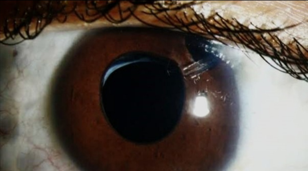
Aphakia = ‘A+phakia’
‘A’ means absence, and ‘Phakic’ refers to a person with an intact natural lens. So Aphakia means absence of the crystalline lens in it’s normal anatomical position of an eye. As well as we can say, the lens been removed or aphakia is a condition where you lose one or both lenses of your eye. It may be considered a condition in which the lens is absent from pupillary area.
○ What about the lens?
》The lens is a clear part of our eye that is found behind our iris (the colored area of our eye). It’s like a camera lens. It is wide and narrow enough to focus the light rays on our retina so that we can see clearly.
■ Common causes of aphakia-
□ Congenital absence of lens. It is a rare condition.
□ Surgical removal of lens,
such as cataract surgery.
□ Posterior dislocation of lens.
□ Heritable disorder associated with dislocation of lens;
▪︎Marfan’s syndrome
▪︎Homocystinuria
▪︎Weill-marchesani syndrome.
□ Aphakia due to absorption of lens matter is noticed rarely after trauma in children.
□ Perforated lesions or ulcers.
□ Congenital anomalies. This leads loss of accommodation, also produces a high degree of hypermetropia and a deep anterior chamber.
□ Traumatic extrusion of lens from the eye also
constitutes a rare cause of aphakia.
□ Posterior dislocation of lens in vitreous causes
optical aphakia.
□ Heritable disorders reported with subluxation of lens;
▪︎Alport’s syndrome
▪︎Aniridia
▪︎Craniofacial dysostosis
▪︎Megalocornea
□ Ocular disorder which can lead to subluxation of lens-
▪︎Buphthalmos
▪︎Exfoliation syndrome
▪︎Intraocular tumor
▪︎Mature or hypermature cataract
□ Retinal detachment, Vitreous detachment and glaucoma are also complications.
▪︎ Aphakic glaucoma: Aphakia causes complex mechanical and biochemical changes in vitreous (a clear gel which fills the space between the lens and retina) and the structure of the anterior part. As aphakic glaucoma, a secondary type of condition that is more difficult to treat than primary glaucoma. People can also acquire glaucoma after cataract surgery, even years the procedure.In patients with aphakia, chronic glaucoma post cataract surgery was found to be less prevalent at 3%.It happens due to the changes in intraocular pressure, which damages the optic nerve.
▪︎ Retinal detachment: After cataract surgery, retinal detachment occurs in 4% of patients. This occurs when the retina becomes detached, causing the eye floaters and a sensation that there is a curtain-like shadow above the eye.
N.B. The risk is higher in young myopic patients.
▪︎ Vitreous detachment: Vitreous is attached to the retina. Cataract surgery can cause significant changes in the vitreous, including detachment from the retina.
● What are the optical changes behind it?
》The lens is important for refraction and therefore the refractive capacity of the eye is significantly reduced as a result of removal.
● Some optical changes occur after removal of crystalline lens-
○ The eye becomes highly hypermetropic.
○ Changes in Cardinal points of the eye –
▪︎ The power of eye decreases from +60D to +44D.
▪︎ The anterior focal point becomes 23.2 mm in front
of the cornea.
▪︎ The posterior focal point lies behind the eyeball.
▪︎ The posterior focal point is about 31 mm behind
the cornea i.e., about 7 mm behind the eyeball.
(The antero-posterior length of eyeball is about
24 mm).
▪︎ There occurs total loss of accommodation.
○ Visual acuity changes in aphakia.
○ Image formation in the aphakic eye.
○ Accommodation in aphakia.
○ Binocular Single Vision in aphakia.
● Types of Aphakia:
As previously we know that some babies are born without eye lenses. This category of aphakia has two types.
● These are,
1. Primary Congenital Aphakia.
2. Secondary Congenital Aphakia.
■ Congenital Primary Aphakia (CPA) – Congenital primary aphakia is a rare eye condition that is present at birth where the lens is absent.
Otherwise, the baby is born without a lens due to genetic mutations or developmental problems.
In some cases, it may be associated with microphthalmia, absence of iris, anterior segment aplasia, and / or other abnormalities of the eye, including sclerocornia (when the cornea merges with the sclera).
N.B. – That primary congenital aphakia can occur when the optic cup has invaginated and partially developed.
■ Secondary Congenital Aphakia – In secondary aphakia, the lens placode has evolved but reproduced before birth (remnants of the lens such as the lens capsule exist).
The development of the lens begins in the third-fourth week of pregnancy(gestation), when the cells on the surface of the optic vesicle thicken and give birth to the optic placode, which turns into the lens vesicle after the invagination process. The latter consists of a single layer of cells covered by a basal lamina, which will eventually form a lens capsule. By the third month, the fibres of the primary lens begin to fill the cavity from the posterior to anterior. This type of aphakia is also associated with exposure to a virus, such as congenital rubella.
Others:
■ Bilateral Congenital Aphakia – There is another type that we can hear, which is bilateral congenital aphakia. A phenomenon of bilateral congenital aphakia is interesting in that both globes developed naturally without evidence of previous inflammation, surgery, or injury.
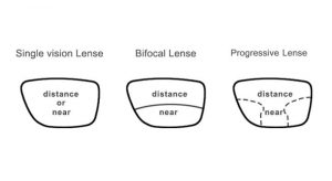
Surgery is the most effective way to treat aphakia in children and adults than using of spectacles. With the help of surgery an artificial intraocular lens (IOL) will insert into the eye which made of silicone, acrylic, or other plastic composition. They are also coated with a special material to help protect your eyes from the sun’s ultraviolet (UV) rays.


