Introduction:
The human eye is a highly evolved structure of our anatomy and has many coexisting and
interdependent elements. It is capable of moving and follow the objects along with accommodating to near and far; the eyes also can see in varying light, and in color. Our two eyes working together give us stereoscopic vision, and depth perception.
The main blood supply of the eye arises from the ophthalmic artery, which gives off orbital and optical group branches. Innervation of the eyeball and surrounding structures is provided by the optic, oculomotor, trochlear, abducens and trigeminal cranial nerves.
Structure and function:
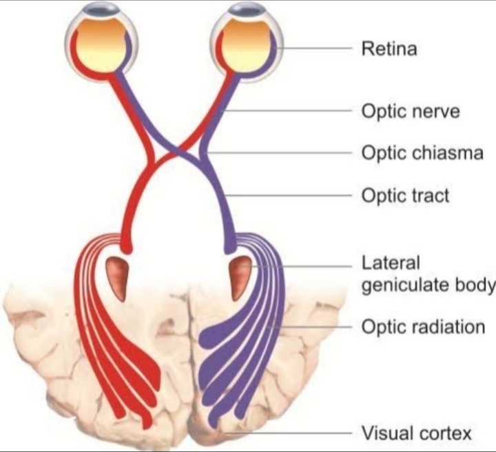
Six cranial nerves innervate motor, sensory, and autonomic structures in the eyes. The six cranial nerves are the optic nerve (CN II), oculomotor nerve (CN III), trochlear nerve (CN IV), trigeminal nerve (CN V), abduces nerve (CN VI), and facial nerve (CN VII). The oculomotor nerve and the trochlear nerve originate in the midbrain. As for the trigeminal nerve, abduces nerve, and facial nerve, they originate from the pons. Interestingly, the optic nerve arises from the optic disc in the eye then enters the brain
as opposed to the other cranial nerve that originates in the brain and then exits peripherally.
Arterial supply:
The branches of the ophthalmic artery comprise the entire arterial supply to the eye. Most commonly the ophthalmic artery branches off the internal carotid artery, distal to the cavernous sinus, then travels through the optic canal. The ophthalmic artery has multiple branches which separate into two
categories: orbital branches and optical branches. The orbital arteries include the ciliary arteries, central retinal artery, and muscular arteries.
Long Posterior Ciliary Arteries: The long posterior ciliary arteries (1 to 2) travel near the optic nerve and pierce the posterior sclera to supply the choroid and ciliary muscle before joining the major arterial
circle of the iris. The major arterial circle of the iris distributes branches to the iris and ciliary body.
Short Posterior Ciliary Arteries: The number of short posterior ciliary arteries vary per individual, often ranging between 6 to 12 arteries that branch off the ophthalmic artery as it crosses the optic nerve medially. These arteries supply the ciliary processes and optic disk. The arterioles branching from the posterior ciliary arteries supply the choroid. The perpendicular terminal arterioles supply choriocapillaris, the blood supply to Bruch’s membrane and outer retina.
Anterior Ciliary Arteries: There are seven anterior ciliary arteries that branch from the muscular arteries and run with the extraocular muscles. The anterior ciliary arteries supply the rectus muscles,
conjunctiva, and sclera before joining the long posterior ciliary arteries to form the major arterial circle of the iris. Each rectus muscle receives its vascular supply from two anterior ciliary arteries, except the
lateral rectus which receives blood supply from only one anterior ciliary artery.
Central Retinal Artery: It is the first branch of the ophthalmic artery. It is a terminal branch supplying the inner layer of the retina, and its occlusion can cause sudden visual loss. It travels inferiorly and within the optic nerve sheath to supply the inner two-thirds of the retina. The artery further divides into superior and inferior arcades, which form the blood-retina barrier.
Muscular branches: The two muscular ranches of the ophthalmic artery that supply extraocular muscles include the medial and lateral muscular branches. The medial artery being larger than the lateral muscular branch.
Vascular supply and drainage :
The arterial input to the eye is provided by several branches from the ophthalmic artery, which is derived from the internal carotid artery in most mammals. These branches include the central retinal artery, the short and long posterior ciliary arteries, and the anterior ciliary arteries. Venous outflow from the eye is
primarily via the vortex veins and the central retinal vein, which merge with the superior and
inferior ophthalmic veins that drain into the cavernous sinus, the pterygoid venous plexus and the facial vein.
The vascular supply of the optic nerve is complex . The optic nerve has three zones referenced to the lamina cribosa, the connective tissue extension of the sclera through which the optic nerve axons and the
central retinal artery and vein pass. The prelaminar (i.e., inside the eye relative to the lamina cribosa) optic nerve is supplied by collaterals from the choroid and retina circulations. The laminar zone is supplied by branches from the short posterior ciliary and pial arteries. The post laminar zone is supplied
by the pial arteries. Venous drainage is via the central retinal vein and pial veins. For the optic
nerve vessels, the laminar zone marks the transition from exposure to the IOP to the cerebral fluid pressure within the optic nerve sheath.
Innervation:
Autonomic innervation of the ocular circulations is restricted to the vessels of the uvea (i.e., the choroid, ciliary body and iris) and optic nerve; the retina appears to lack sympathetic and parasympathetic nerves7–9. The postganglionic sympathetic nerves originate in the superior cervical ganglion. Parasympathetic innervation originates in the pterygopalatine ganglion via the facial nerve.


