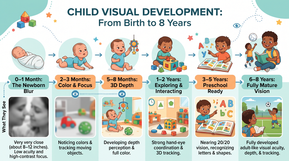
Every time I hand a tiny pair of pediatric frames to a parent in my optometry clinic, I hear the same question: “what does the world actually look like through my baby’s eyes?” Most people imagine human eyes work like smartphone cameras, ready to capture crisp images the moment a newborn opens their lids. In reality, birth is only the opening chapter of an eight-year optical and neurological construction project. Whether you are a fellow clinician, an optometrist or ophthalmologist or a parent gazing at your newborn, the journey from blurry shadows to three-dimensional sight is an extraordinary biological story.
Month One: A Misty World
When a full-term newborn opens their eyes, the room looks like a soft watercolor painting. Clinically, preferential-looking tests show that neonatal grating acuity starts around 1 cycle per degree (cpd), equivalent to roughly 20/600 (6/180) Snellen or 1.48 logMAR (Leat et al., 2009). Why is early sight so hazy? The answer lies at the center of the retina: the fovea. Histological studies reveal that although the foveal pit begins forming around fetal week 25 as inner retinal layers migrate outward, a newborn’s rod-free foveola still spans over 1,000 microns and holds a single layer of short, wide cones measuring 7.5 microns across with outer segments 5 to 14 microns long (Hendrickson et al., 2012; Yuodelis & Hendrickson, 1986).
Because these cones are loosely packed, 18 cones per 100 microns at one week postnatal, the infant eye has a wide depth of focus (Banks, 1980; Yuodelis & Hendrickson, 1986). Dynamic retinoscopy shows that one-month-olds rarely accommodate accurately across distances, not from ciliary muscle weakness, but because coarse retinal resolution fails to trigger fine focus adjustments (Banks, 1980). Despite the myth that newborns see only in black and white, over 75% of neonates orient toward large, saturated red patches against gray, though over 80% miss blue targets (Skelton et al., 2022). Up close (8 to 12 inches away) high-contrast contours dominate their view.
Months Two to Six: Awakening
Between two and six months, visual development accelerates. Inside the macula, cones migrate inward toward the foveal pit and narrow, tightening the sensory feedback loop driving accommodation (Banks, 1980; Hendrickson et al., 2012). By three months, infants maintain accurate accommodation across moderate hyperopic demands (Mutti et al., 2009). Simultaneously, color perception blossoms. While the red–green (L- and M-cone opponent) channel operates first, the short-wavelength blue–yellow (S-cone) mechanism matures four to eight weeks later, making infants trichromatic by three months and able to group hues into categories like red, green, blue, and yellow by four months (Skelton et al., 2022).
Around month four comes a milestone parents love watching—when a baby reaches out and snatches a dangling toy. That leap relies on stereopsis, or binocular 3D depth perception. Unlike visual acuity’s gradual climb, binocular disparity tuning in the primary visual cortex (V1) switches on abruptly (Held et al., 1980). Stereopsis emerges at a mean age of 16 weeks and reaches 1 minute of arc (60 arcseconds) or better by 21 weeks (Held et al., 1980). Meanwhile, Teller Acuity Cards show grating acuity improving from 1.31 logMAR (20/400) at three months to 0.97 logMAR (20/185) at nine months, while visual evoked potentials (VEP) approach 20/20 cortical resolution by six months (American Optometric Association, 2017; Leat et al., 2009; Mutti et al., 2009; Teller et al., 1986).
Infancy to Preschool: Optical Tuning
Why do most babies outgrow early farsightedness without glasses? Term newborns have axially short eyes, averaging +1.00 D to +2.50 D of cycloplegic hyperopia, while 30% to 50% have ≥ 1.00 D of astigmatism (American Optometric Association, 2017; Mutti et al., 2009). During the first 18 months the eye undergoes emmetropization, matching axial growth to optical power in proportion to initial refractive error and accommodation (Mutti et al., 2009). In the Berkeley Infant Biometry Study, mean cycloplegic hyperopia fell from +2.13 D at three months to +1.32 D at nine months and +1.10 D at 18 months (0.85 logMAR acuity), while astigmatism stabilizes by ages 2.5 to 3 years (American Optometric Association, 2017; Mutti et al., 2009).
Microscopically, foveal architecture keeps remodeling. By 15 months, inner retinal cells finish clearing the foveal pit, and elongating cone axons (fibers of Henle) thicken the central outer plexiform layer (Hendrickson et al., 2012). By 45 months, central cones slim from 7.5 microns to their adult 2-micron width and narrow the foveola to 650–700 microns, though cone density at age 3.8 years (108,400 cones/mm²) remains half the adult peak of 208,200 cones/mm² (Hendrickson et al., 2012; Yuodelis & Hendrickson, 1986).
Ages Three to Eight: Plasticity
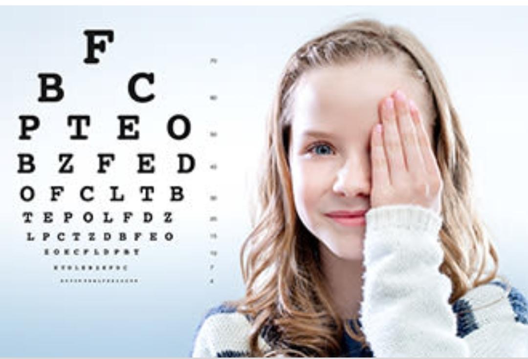
In preschool, testing shifts from gratings to symmetric matching optotypes with crowding bars, such as Lea Symbols or crowded HOTV charts (National Research Council, 2002). Healthy 3 to 4-year-olds pass the 20/40 (0.30 logMAR) line, reach 20/25 (0.10 logMAR) on logMAR charts by ages 5.5 to 7, and achieve adult-like 20/20 (0.00 logMAR) acuity between ages 6 and 8, while contrast sensitivity refines into adolescence (Leat et al., 2009; National Research Council, 2002).
From birth to age seven or eight, the brain traverses its visual critical period (Daw, 1998; Hensch & Quinlan, 2018). If one eye is blurred by anisometropia or hyperopia (≥ +2.00 D) or misaligned by strabismus, V1 ocular dominance shifts away from the weaker eye, causing amblyopia—the leading cause of childhood monocular vision loss (American Optometric Association, 2017; Daw, 1998). Around ages seven to eight, parvalbumin inhibitory circuits and molecular “brakes” like perineuronal nets and Lynx1 proteins stabilize synapses, making amblyopia harder to reverse (American Optometric Association, 2017; Hensch & Quinlan, 2018).
Exam Milestones and Red Flags
Because 1 in 5 preschoolers has a vision disorder, screening before the critical period closes is vital (American Optometric Association, 2017):
• Infant exam (6–12 months): Evaluates cycloplegic refraction, emmetropization, motility, and ocular health.
• Preschool exam (3–5 years): Detects amblyogenic anisometropia, hyperopia, or esotropia while plasticity peaks.
• School-entry exam (ages 5–6 and annually): Confirms 20/20 acuity, convergence, and accommodation.
• Parent red flags: Seek care for a constant eye turn (strabismus) or drifting past four months, a white pupil reflex in flash photos (leukocoria, warning of cataract or retinoblastoma), rhythmic eye jerking (nystagmus), a drooping lid (ptosis), squinting, or failure to fixate and follow by three months (American Optometric Association, 2017; National Research Council, 2002).
References
• Leat, S. J., Yadav, N. K., & Irving, E. L. (2009). Development of visual acuity and contrast sensitivity in children. Journal of Optometry, 2(1), 19–26. https://pmc.ncbi.nlm.nih.gov/articles/PMC3972638/
• Hendrickson, A., Possin, D., Vajzovic, L., & Toth, C. (2012). Histological development of the human fovea from midgestation to maturity. American Journal of Ophthalmology, 154(5), 767–778.e2. https://pmc.ncbi.nlm.nih.gov/articles/PMC3509500/
• Yuodelis, C., & Hendrickson, A. (1986). A qualitative and quantitative analysis of the human fovea during development. Vision Research, 26(6), 847–855. https://pubmed.ncbi.nlm.nih.gov/3750868/
• Banks, M. S. (1980). The development of visual accommodation during early infancy. Child Development, 51(3), 646–666. https://pubmed.ncbi.nlm.nih.gov/7418504/
• Skelton, A. E., Maule, J., & Franklin, A. (2022). Infant color perception: Insight into perceptual development. Child Development Perspectives, 16(2), 90–95. https://pmc.ncbi.nlm.nih.gov/articles/PMC9314692/
• Mutti, D. O., Mitchell, G. L., Jones, L. A., Friedman, N. E., Frane, S. L., Lin, W. K., Moeschberger, M. L., & Zadnik, K. (2009). Accommodation, acuity, and their relationship to emmetropization in infants. Optometry and Vision Science, 86(6), 666–676. https://pmc.ncbi.nlm.nih.gov/articles/PMC2706284/
• Held, R., Birch, E., & Gwiazda, J. (1980). Stereoacuity of human infants. Proceedings of the National Academy of Sciences, 77(9), 5572–5574. https://pubmed.ncbi.nlm.nih.gov/6933571/
• American Optometric Association. (2017). Evidence-based clinical practice guideline: Comprehensive pediatric eye and vision examination. https://www.aoa.org/aoa/documents/practice%20management/clinical%20guidelines/ebo%20guidelines/comprehensive%20pediatric%20eye%20and%20vision%20exam.pdf
• Teller, D. Y., McDonald, M. A., Preston, K., Sebris, S. L., & Dobson, V. (1986). Assessment of visual acuity in infants and children: The acuity card procedure. Developmental Medicine & Child Neurology, 28(6), 779–789. https://pubmed.ncbi.nlm.nih.gov/3817317/
• National Research Council. (2002). Assessment of vision in infants and children. In P. Lennie & S. B. Van Hemel (Eds.), Visual impairments: Determining eligibility for Social Security benefits (pp. 163–205). National Academies Press. https://www.ncbi.nlm.nih.gov/books/NBK207548/
• Daw, N. W. (1998). Critical periods and amblyopia. Archives of Ophthalmology, 116(4), 502–505. https://pubmed.ncbi.nlm.nih.gov/9565050/
• Hensch, T. K., & Quinlan, E. M. (2018). Critical periods in amblyopia. Visual Neuroscience, 35, Article E014. https://pmc.ncbi.nlm.nih.gov/articles/PMC6047524/