
Introduction
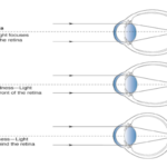
Myopia, or nearsightedness, is a refractive condition characterised by the elongation of
the eyeball, causing distant objects to appear blurred as light rays focus anterior to the
retina . While traditionally considered a benign and easily correctable condition, myopia
has now emerged as a major global public health concern due to its rapidly increasing
prevalence and its association with sight-threatening complications in high and
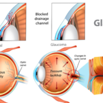
pathologic forms . These include myopic maculopathy, retinal detachment, glaucoma,
and choroidal neovascularization, which significantly contribute to irreversible vision loss
The terminology itself reflects this evolution. The International Myopia Institute (IMI)
distinguishes myopia correction restoring clear vision from interventions intended to
slow progression. Myopia management encompasses a broader, longitudinal approach
involving risk assessment, intervention and monitoring.
This is particularly relevant because the projected global burden of myopia is
substantial. Previous epidemiological modelling estimated that approximately 49.8% of
the world’s population could be myopic by 2050, with nearly 1 billion people potentially
affected by high myopia.
The clinical concern is not simply the number of individuals requiring optical correction.
Increasing axial length and higher degrees of myopia are associated with a greater
lifetime burden of ocular pathology, making prevention of excessive axial elongation an
important public-health and clinical objective.
Why Axial Length Deserves a Central Role
Spherical equivalent refraction remains fundamental to diagnosis and clinical decision-
making, but it does not directly measure ocular growth.
Axial length provides a structural parameter that can be followed longitudinally. Modern
optical biometers allow clinicians to measure axial length with high precision, making
repeated measurements particularly valuable in children undergoing myopia
management.
The distinction becomes clearer when treatment efficacy is considered.
The 2025 IMI intervention review recommends interpreting efficacy primarily through
slowing of axial elongation, alongside refractive progression. Its evidence synthesis
included randomized controlled trials with at least 12 months of follow-up and axial-
length measurements for established interventions.
Epidemiology and global burden of myopia
The predominant cause of myopic progression is excessive axial elongation. Visual
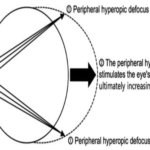
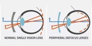
signals, particularly peripheral hyperopic defocus—where light focuses behind the
peripheral retina—trigger compensatory mechanisms that stimulate ocular growth. This
phenomenon is mediated by neuromodulators such as dopamine, nitric oxide, and
retinoic acid, which influence biochemical pathways in the retina and sclera . These
signals alter scleral extracellular matrix remodelling, leading to thinning and elongation
of the posterior globe. Experimental evidence from animal models supports the theory
that manipulating peripheral retinal image quality can directly influence axial length
growth .
Advances in imaging modalities, such as optical coherence tomography (OCT) and
magnetic resonance imaging (MRI), have revealed key structural changes associated
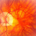
with progressive and pathological myopia. These include scleral thinning, choroidal
atrophy, and the development of posterior staphyloma . High and pathologic myopia are
also associated with tractional complications, such as myopic macular degeneration and
foveoschisis, which contribute to permanent vision loss in advanced stages.
Classification of myopia can be approached from several clinical angles. Based on
refractive severity, myopia is typically categorised as low (<–3.00 dioptres), moderate
(–3.00 to –6.00 dioptres), and high (>–6.00 dioptres) . Pathologic myopia, however,
refers to cases where degenerative structural changes in the posterior segment are
present, regardless of refractive error magnitude. Anatomically, myopia is divided into
axial myopia—driven by globe elongation—and refractive myopia, which results from
increased curvature or refractive index of the cornea or lens . Axial myopia is the most
prevalent and carries a greater risk of progression and associated pathology.
Age of onset further stratifies myopia into congenital, childhood or youth-onset, early
adult-onset, and late-onset categories. Congenital myopia is often linked with systemic
conditions or genetic syndromes. Youth-onset myopia typically manifests during school-
age years and is strongly associated with environmental risk factors such as excessive
near work and limited outdoor activity. Late-onset myopia is more commonly linked to
occupational visual demands and may also result from nuclear sclerosis in aging lenses.
What Do the Numbers Tell Us?
The magnitude of treatment effects can appear deceptively small because axial length
changes are measured in millimetres.
According to the 2025 IMI evidence synthesis, orthokeratology demonstrated a median
reduction in axial elongation of approximately 0.17 mm at one year and 0.30 mm at two
years. Soft multifocal/contact-lens approaches demonstrated efficacy of up to
approximately 0.19 mm at one year and 0.28 mm at three years, while newer myopia-
control spectacle designs demonstrated efficacy of up to approximately 0.35 mm over
two years.
For atropine, the median axial-length efficacy reported for 0.01% atropine was
approximately 0.08 mm at one year and 0.12 mm at two years. Higher concentrations
have demonstrated greater efficacy in some randomized trials, with two-year efficacy
reaching approximately 0.50 mm in the IMI review.
These figures should not be interpreted as interchangeable outcomes between different
trials. Study populations, baseline age, myopia level, follow-up duration, adherence and
control groups differ. Nevertheless, they illustrate an important principle:
In myopia management, a fraction of a millimetre can represent a clinically meaningful
difference in ocular growth trajectory.
The LAMP Study: Why Concentration Matters
One of the most influential datasets in pharmacological myopia management is the
Low-Concentration Atropine for Myopia Progression (LAMP) study.
The randomized, double-masked trial compared 0.05%, 0.025% and 0.01% atropine
with placebo in children aged 4–12 years.
After one year, mean axial elongation was:
0.20 ± 0.25 mm with 0.05% atropine
0.29 ± 0.20 mm with 0.025%
0.36 ± 0.29 mm with 0.01%
0.41 ± 0.22 mm with placebo
The response therefore demonstrated a concentration-dependent pattern.
The findings are particularly important because they demonstrate why refractive
progression alone can be insufficient when evaluating treatment response. The ocular
biometric data indicate that low-concentration atropine’s antimyopic effect is
substantially reflected through reduced axial elongation.
Long-Term Treatment: The Five-Year LAMP Perspective
The longer-term LAMP data provide an even more important clinical lesson: myopia
management is not necessarily a short-duration intervention.
In the five-year follow-up, cumulative spherical-equivalent progression among children
who continued treatment was approximately:
-1.34 D with initial 0.05% atropine
-1.97 D with initial 0.025% atropine
-2.34 D with initial 0.01% atropine
The study also reported significant differences in axial-length elongation between
treatment groups.
Treatment cessation also deserves attention. Among children assigned to a pro re nata
retreatment strategy after treatment cessation, 87.9% required retreatment during the
relevant follow-up period.
This does not mean every child requires lifelong atropine. Rather, it emphasizes the
importance of continued surveillance after treatment modification or cessation.
Rebound: A Clinical Variable, Not a Footnote
The concept of rebound is another reason why treatment decisions should be
longitudinal.
In the three-year LAMP analysis, children who stopped atropine experienced faster axial
elongation during the washout year than those who continued treatment. For example,
in the 0.05% group, axial elongation during year three was approximately 0.33 mm after
washout versus 0.17 mm with continued treatment.
However, the authors also reported that differences in rebound between concentrations
were clinically small, and older age at cessation and lower treatment concentration were
associated with smaller rebound effects.
The practical message is clear: stopping therapy should be treated as a monitored
clinical decision rather than an automatic endpoint.
The Axial-Length-Centred Clinical Examination
A contemporary myopia-management examination should therefore extend beyond a
conventional refraction.
A useful longitudinal dataset may include:
1. Cycloplegic spherical equivalent
Provides a reliable measure of refractive status, particularly in children.
2. Axial length
Allows direct monitoring of ocular elongation.
3. Best-corrected visual acuity
Confirms functional visual performance.
4. Binocular vision and accommodative assessment
Relevant when evaluating the broader visual environment and treatment response.
5. Ocular health examination
Essential for identifying pathology and establishing treatment safety.
6. Age of onset and progression history
Earlier onset and rapid progression generally warrant closer attention.
7. Family history and environmental factors
Parental myopia and visual/environmental exposure contribute to risk assessment.
8. Treatment adherence
A poor response may represent inadequate biological efficacy or simply inadequate
treatment exposure.
9. Optical
Spectacle lenses
D.I.MS (MiYOSMART by Hoya)
H.A.L.T (Stellest lenses by Essilor)
C.A.R.E (MyoCare by Zeiss)
D.O.T (diffusion Optics Technology)
Multifocal contact lenses
Ortho-K lenses
10. Red light therapy
Repeated low-level red-light therapy (≈650 nm) has shown promising potential in
slowing axial elongation and myopia progression.
Its role is emerging, with further long-term evidence needed to establish optimal
protocols and safety.
This approach converts isolated clinical measurements into a patient-specific growth
trajectory.
The Future: Predictive and Personalized Myopia Care
The next phase of myopia management will likely move toward increasingly
individualized treatment algorithms. The IMI’s 2025 instrumentation update highlights
developments in axial-length measurement, imaging, aberrometry and emerging AI-
based prediction tools.
The objective is not simply to identify children who are already progressing, but
potentially to identify which eyes are most likely to progress rapidly and which
intervention is most appropriate for that individual.
Ultimately, the success of myopia management should not be measured solely by how
many dioptres of spectacle power have been avoided.
It should be measured by how effectively we can alter the trajectory of ocular growth.