■ Introduction
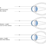
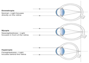
Myopia is traditionally understood as a refractive
condition in which distant objects appear blurred
because incoming light focuses in front of the retina.
However, myopia is not simply a matter of refractive
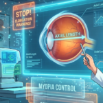
power. In eyes with excessive axial elongation,
progressive structural changes can develop in the
posterior segment, affecting the retina, retinal
pigment epithelium (RPE), choroid, Bruch’s
membrane, sclera and optic nerve.
When excessive axial elongation produces
characteristic pathological changes in the posterior
eye, the condition is referred to as pathologic
myopia.
Pathologic myopia is clinically important because its
complications can result in progressive and
sometimes irreversible visual impairment.
Importantly, high myopia and pathologic myopia are
not synonymous. High myopia describes a high
degree of myopic refractive error, whereas
pathologic myopia is defined by the presence of
characteristic structural complications, such as
posterior staphyloma or myopic maculopathy. These
pathological changes can occasionally occur even in
eyes that do not meet conventional high-myopia
refractive thresholds.
1. What Is Pathologic Myopia?
The International Myopia Institute defines pathologic
myopia as:
– Excessive axial elongation associated with myopia.
– Structural changes occurring in the posterior
segment of the eye.
– Typical manifestations include:
– Posterior staphyloma
– Myopic maculopathy
– Myopia-associated optic neuropathy
– These changes may ultimately cause loss of best-
corrected visual acuity.
This distinction is clinically important.
● High Myopia vs Pathologic Myopia
● High myopia:
– Generally refers to a high degree of refractive
myopia.
– A commonly used research definition is spherical
equivalent of ≤ −6.00 D.
– It does not automatically mean that pathological
retinal changes are present.
● Pathologic myopia:
– Is characterized by structural damage or
pathological changes in the posterior segment.
– It may occur with high myopia but is not defined
solely by refractive power.
– Posterior staphyloma and myopic maculopathy are
important clinical indicators.
2. Why Does Pathologic Myopia Develop?
The central factor is excessive axial elongation. As
the eye becomes progressively elongated:
– The posterior sclera undergoes biomechanical
expansion.
– The choroid becomes progressively thinner.
– The RPE and Bruch’s membrane are subjected to
mechanical stretching.
– The retina may become progressively thinner.
– The relationship between the retina, choroid and
underlying sclera changes.
– Abnormal curvature of the posterior globe can
produce a posterior staphyloma.
These changes create an environment in which
multiple retinal and choroidal complications can
develop.
The process is therefore not simply:
More minus power → worse vision
3. Posterior Staphyloma: A Major Structural
Feature
One of the most characteristic features associated
with pathologic myopia is posterior staphyloma. It
refers to an outward protrusion or ectatic
deformation of the posterior wall of the eye, usually
involving the posterior pole.
● Why is it important?
● Posterior staphyloma can:
– Alter the normal contour of the posterior globe.
– Increase mechanical stress on the retina and
choroid.
– Contribute to chorioretinal atrophy.
– Be associated with myopic traction maculopathy.
– Influence the development and progression of
macular complications.
Modern wide-field imaging and three-dimensional
imaging techniques have improved the ability to
identify and characterize staphyloma.
4. Myopic Maculopathy
Myopic maculopathy represents one of the most
important causes of vision loss associated with
pathologic myopia. It encompasses a spectrum of
progressive structural abnormalities involving the
macula.
● Important manifestations include:
– Diffuse chorioretinal atrophy
– Patchy chorioretinal atrophy
– Lacquer cracks
– Myopic macular neovascularization
– CNV-related macular atrophy
– Fuchs spot
– Myopic traction maculopathy
– Macular hole
– Foveal detachment
The severity of myopic maculopathy can range from
relatively subtle changes to extensive macular
destruction with significant visual impairment.
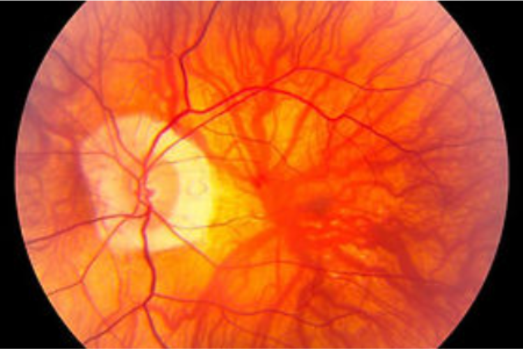
5. Diffuse Chorioretinal Atrophy
Diffuse chorioretinal atrophy is an important
manifestation of myopic maculopathy. It involves
generalized thinning and attenuation of the:
– Choroid
– RPE
– Choriocapillaris
– Associated retinal structures
Clinically, the fundus may demonstrate a more
visible underlying choroidal vasculature because of
thinning of the RPE and choroid.
● Why does it matter?
● Progressive chorioretinal thinning can lead to:
– Reduced photoreceptor support
– Progressive macular dysfunction
– Reduced visual acuity
– Increased susceptibility to more advanced atrophic
changes
Diffuse atrophy can therefore represent an important
stage in the continuum of myopic macular
degeneration.
6. Patchy Chorioretinal Atrophy
Patchy atrophy represents a more localized and
severe form of chorioretinal tissue loss. These areas
may appear as sharply demarcated regions where
the RPE and choroid are markedly attenuated.
● Patchy atrophy can:
– Involve the macula.
– Enlarge over time.
– Directly affect photoreceptor integrity.
– Cause permanent visual field or acuity loss
depending on its location.
Recent longitudinal imaging studies have
demonstrated that enlargement of patchy atrophy
can be an important pattern of progression in highly
myopic eyes.
7. Lacquer Cracks: Mechanical Damage to Bruch’s
Membrane
Lacquer cracks are yellowish linear lesions
commonly associated with pathologic myopia. They
represent breaks or mechanical disruptions involving
Bruch’s membrane, usually occurring as the
posterior segment undergoes progressive stretching.
● Clinical significance :-
● Lacquer cracks are important because they can:
– Represent structural damage caused by
progressive ocular stretching.
– Become associated with secondary vascular
complications.
– Provide a pathway for abnormal choroidal vessels
to develop beneath or through the damaged tissue.
Patients with lacquer cracks therefore require careful
macular evaluation and longitudinal monitoring.
8. Myopic Macular Neovascularization
One of the most vision-threatening complications of
pathologic myopia is myopic macular
neovascularization (MNV). Mechanical disruption of
Bruch’s membrane and changes in the choroid-RPE
complex may facilitate the development of abnormal
neovascular tissue.
● These abnormal vessels can cause:
– Subretinal hemorrhage
– Exudation
– Macular structural disruption
– Sudden or progressive visual deterioration
A patient with pathologic myopia who develops new
central distortion, reduced vision or a new scotoma
requires prompt macular assessment.
● Management :-
Anti-VEGF therapy has become an important
treatment for myopic macular neovascularization.
Early detection is critical because untreated
neovascular activity may result in permanent
macular damage and subsequent macular atrophy.
9. CNV-Related Macular Atrophy
Even after an episode of myopic macular
neovascularization becomes inactive, structural
damage may remain.
● The affected region may subsequently develop:
– RPE atrophy
– Choriocapillaris loss
– Photoreceptor damage
– Macular scar formation
– Permanent reduction in visual function
Therefore, successful suppression of neovascular
activity does not necessarily mean complete
restoration of the macula. Long-term monitoring
remains important because the visual consequences
of previous MNV can persist or evolve.
10. Myopic Traction Maculopathy
Pathologic myopia can also produce mechanical
abnormalities at the macula. Myopic traction
maculopathy (MTM) includes a spectrum of traction-
related changes associated with excessive axial
elongation and posterior globe deformation.
● Possible findings include:
– Retinoschisis
– Foveal detachment
– Epiretinal membrane-related changes
– Macular hole
– Combined tractional abnormalities
Optical coherence tomography (OCT) is particularly
valuable because many of these abnormalities may
not be fully appreciated on routine fundus
examination.
11. Macular Hole and Foveal Detachment
Advanced mechanical changes can result in the
development of a full-thickness macular hole or
foveal detachment.
● The combination of:
– Posterior staphyloma
– Vitreoretinal traction
– Retinal stretching
– Progressive axial elongation
Can create substantial mechanical stress at the
fovea.
● These complications may produce:
– Central visual distortion
– Reduced visual acuity
– Central scotoma
– Difficulty with reading and fine visual tasks
Some advanced cases may require vitreoretinal
surgical intervention.
12. Choroidal Thinning
Choroidal thinning is one of the key structural
features of pathologic myopia. Progressive axial
elongation is associated with reduction in choroidal
thickness, particularly in the posterior pole.
● The choroid plays an important role in:
– Supplying oxygen and nutrients to the outer retina.
– Supporting the RPE and photoreceptors.
– Maintaining the metabolic environment of the
macula.
Therefore, severe choroidal thinning may contribute
to the development and progression of myopic
macular pathology.
OCT and enhanced-depth imaging can provide
valuable information about these changes.
13. Peripheral Retinal Complications
Although the macula receives considerable attention
in pathologic myopia, the peripheral retina should
not be overlooked.
● Highly myopic eyes may have an increased
susceptibility to:
– Retinal breaks
– Retinal tears
– Rhegmatogenous retinal detachment
– Peripheral retinal degeneration
The combination of a long axial length, peripheral
retinal changes and vitreous alterations can increase
the risk of retinal complications.
● Warning symptoms
Patients should be educated to seek urgent
ophthalmic evaluation if they experience:
– Sudden onset or marked increase in floaters
– Flashes of light
– A curtain or shadow over the visual field
– Sudden reduction in vision
These symptoms can indicate a retinal tear or retinal
detachment and should not be ignored.
14. Optic Nerve and Peripapillary Changes
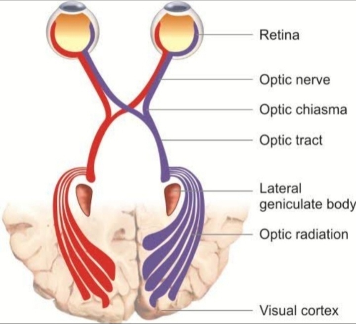
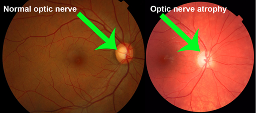
Pathologic myopia can also affect the optic nerve
and peripapillary region.
● Common structural findings include:
– Tilted optic disc
– Peripapillary atrophy
– Enlarged peripapillary zones
– Changes in the optic nerve head configuration
– Myopia-associated glaucoma-like optic neuropathy
These anatomical changes can make interpretation
of the optic disc and OCT retinal nerve fibre layer
more challenging.
● A highly myopic optic nerve should therefore be
interpreted carefully and in combination with:
– Intraocular pressure
– Visual field findings
– Optic nerve appearance
– Macular ganglion cell analysis
– Longitudinal structural changes
15. The Role of OCT in Pathologic Myopia
Optical coherence tomography has transformed the
evaluation of pathologic myopia. OCT can detect
structural abnormalities that may be difficult to
identify clinically.
● OCT can help evaluate:
– Choroidal thickness
– Retinal thinning
– Retinoschisis
– Foveal detachment
– Macular holes
– Myopic traction maculopathy
– Macular neovascularization
– RPE alterations
– Photoreceptor disruption
– Atrophic changes
Serial OCT examinations are particularly useful
because they allow clinicians to compare structural
changes over time rather than relying solely on a
single examination.
16. Fundus Photography and Multimodal
Imaging
A comprehensive assessment may require more
than one imaging modality.
● Depending on the clinical presentation, evaluation
can include:
– Dilated fundus examination
– Colour fundus photography
– OCT
– OCT angiography
– Fundus autofluorescence
– Ultra-widefield imaging
– Fluorescein angiography when indicated
– Indocyanine green angiography in selected cases
Each modality provides different information.
● For example:
Fundus photography
– Documents macular and peripheral changes.
– Useful for longitudinal comparison.
□ OCT
– Demonstrates cross-sectional retinal and
choroidal anatomy.
□ OCT-A
– Helps evaluate retinal and choroidal vascular
networks without dye injection.
□ Fundus autofluorescence
– Can provide information about RPE-related
abnormalities.
A multimodal approach can therefore improve
detection and characterization of pathologic
changes.
17. Classification of Myopic Maculopathy
The META-PM classification system is widely used
to describe myopic maculopathy based on fundus
appearance.
● It includes categories progressing from:
– No myopic maculopathy
– Tessellated fundus
– Diffuse chorioretinal atrophy
– Patchy chorioretinal atrophy
– Macular atrophy
● Additional “plus lesions” include important
complications such as:
– Lacquer cracks
– Myopic choroidal neovascularization
– Fuchs spot
More recent approaches increasingly incorporate
OCT-based structural information, particularly for
conditions such as myopic traction maculopathy and
dome-shaped macula.
18. Why Regular Monitoring Matters
Pathologic myopia can progress even when the
refractive prescription appears relatively stable.
Therefore, clinical monitoring should not focus
exclusively on refraction.
● A comprehensive follow-up may include:
– Best-corrected visual acuity
– Refraction
– Axial length assessment when appropriate
– Dilated retinal examination
– Fundus photography
– OCT
– Peripheral retinal evaluation
– Assessment of new symptoms
– Comparison with previous imaging
The frequency of follow-up should be individualized
according to age, severity, previous complications,
axial length, symptoms and documented structural
progression.
19. Patient Education: An Essential Component
Patients with high or pathologic myopia should
understand that clear vision with spectacles does
not necessarily mean that the eye is structurally
normal.
● Important counselling points include:
– Myopia can be associated with long-term retinal
and choroidal complications.
– Structural changes may progress even when
refractive power changes little.
– Regular retinal evaluation is important.
– Sudden flashes, floaters or a curtain-like shadow
require urgent evaluation.
– New distortion or sudden central vision changes
should not be ignored.
– Previous myopic complications may require lifelong
monitoring.
Patient education can therefore play an important
role in preventing delayed presentation of sight-
threatening complications.
20. Prevention and Myopia Management
Not every patient with myopia will develop
pathologic myopia. However, reducing excessive
myopic progression, particularly during childhood
and adolescence, is an important public-health and
clinical objective.
● Evidence-based myopia management may include
appropriate:
– Optical interventions
– Pharmacological interventions
– Behavioural approaches
– Increased outdoor exposure
– Regular monitoring of refractive error and axial
length where appropriate
The long-term relationship between childhood
myopia-control interventions and prevention of adult
pathologic complications continues to be studied.
Nevertheless, controlling excessive myopic
progression remains an important strategy in
reducing the future burden associated with high and
pathologic myopia.
■ Conclusion
Pathologic myopia represents far more than a high
spectacle prescription. It is a structural disease of
the elongated eye, in which progressive changes
involving the sclera, choroid, Bruch’s membrane,
RPE, retina and optic nerve can lead to significant
visual morbidity.
● The chorioretinal complications form a broad
spectrum:
– Diffuse chorioretinal atrophy
– Patchy chorioretinal atrophy
– Lacquer cracks
– Myopic macular neovascularization
– CNV-related macular atrophy
– Myopic traction maculopathy
– Macular hole
– Foveal detachment
– Posterior staphyloma-associated macular changes
The clinical challenge is that these complications
may evolve gradually, remain asymptomatic in their
early stages, or produce irreversible structural
damage before significant visual symptoms become
apparent. Modern multimodal imaging—particularly
OCT, OCT angiography, fundus photography and
wide-field imaging—has significantly improved the
ability to identify and monitor these changes.
Ultimately, the management of myopia should
extend beyond achieving clear distance vision. The
long-term goal is to recognize eyes at risk, identify
pathological changes early, monitor structural
progression and provide timely intervention when
sight-threatening complications develop.