Introduction
Every minute, somewhere in the developing world, a child loses their sight — not from
trauma, not from infection alone, but from a deficiency of a single, inexpensive
micronutrient: Vitamin A. Vitamin A deficiency (VAD) remains one of the most
consequential and yet most preventable nutritional disorders of the 21st century.
Defined clinically as a serum retinol concentration below 0.70 µmol/L, VAD affects an
estimated 190 million preschool-aged children and 19 million pregnant women globally,
with the burden concentrated overwhelmingly in sub-Saharan Africa and South and
Southeast Asia (World Health Organization [WHO], 2009).
The consequences of VAD extend far beyond the eye. It impairs immune function,
increases childhood mortality, and disrupts normal growth and development. Yet the
ocular manifestations — collectively termed xerophthalmia — represent its most visible
and devastating expression. VAD is the leading nutritional cause of preventable
childhood blindness, responsible for an estimated 250,000 to 500,000 children losing
their sight annually, half of whom die within twelve months of going blind (Sommer,
2008).
Despite decades of intervention, VAD persists as a stubborn public health challenge.
Biochemical Mechanisms of Vitamin’s Role in Vision
Vitamin A — the umbrella term for retinol, retinal, retinoic acid, and provitamin A
carotenoids — plays an irreplaceable role in the visual cycle. The primary mechanism is
the photochemical conversion of 11-cis-retinal, a form of vitamin A aldehyde, with the
protein opsin to form rhodopsin in the rod photoreceptors of the retina (Tanumihardjo,
2011). Rhodopsin is the photopigment responsible for low-light, scotopic vision. Upon
absorbing a photon of light, 11-cis-retinal isomerizes to all-trans-retinal, triggering a
conformational change that initiates the phototransduction cascade — ultimately
generating a nerve impulse that the brain interprets as vision (Rando, 1990).
In conditions of VAD, the supply of 11-cis-retinal to the outer segments of rod
photoreceptors becomes depleted. As rhodopsin cannot be regenerated efficiently, rod
sensitivity diminishes in a dose-dependent manner. This produces the earliest and most
diagnostically sensitive sign of VAD: night blindness, or nyctalopia. Beyond the retina,
retinoic acid — another bioactive form of vitamin A — regulates the differentiation and
maintenance of epithelial tissues throughout the body, including the conjunctival goblet
cells that produce the mucin layer of the tear film (Sommer, 2008). Without adequate
retinoic acid signaling, goblet cell density declines, the tear film becomes unstable, and
the ocular surface begins to keratinize — a process that culminates in xerophthalmia.
Ocular Conditions Caused by Vitamin A Deficiency
VAD produces a spectrum of ocular manifestations that progress in severity, collectively
classified under the WHO grading system for xerophthalmia:
Night blindness (XN) is the earliest functional sign and is reported subjectively by
patients as difficulty seeing in dim light. It reflects depleted retinal rhodopsin and is
reversible with prompt supplementation. In clinical field studies in Nepal and
Bangladesh, maternal night blindness during pregnancy has been used as a cost-
effective, population-level indicator of VAD severity (West, 2003).
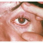
Conjunctival xerosis (X1A) represents drying and wrinkling of the bulbar conjunctiva
due to loss of goblet cell density and mucin secretion. Bitot's spots (X1B) — foamy,
cheese-like plaques on the temporal conjunctiva — are pathognomonic of VAD and are
caused by accumulation of keratinized epithelial debris. Although Bitot's spots may
persist in older children even after vitamin A status is restored, their presence in
children under five years is strongly indicative of active deficiency (Sherwin et al., 2012).
Corneal xerosis (X2) and corneal ulceration (X3A) represent the most vision-
threatening stages of xerophthalmia. The desiccated, keratinized corneal surface
becomes vulnerable to secondary bacterial and viral infections, and even minor trauma
can precipitate ulceration (Sommer & West, 1996). Corneal melt — keratomalacia (X3B)
— is the most severe manifestation, in which rapid liquefactive necrosis of the corneal
stroma occurs, often bilaterally. This process is devastatingly swift: a child may progress
from early xerosis to total corneal destruction within days during a febrile illness such as measles, when metabolic demand for vitamin A spikes and existing stores are rapidly depleted (Sommer, 2008).
Corneal scarring (XS), the sequela of healed ulceration or keratomalacia, results in
permanent visual impairment or total blindness. Unlike earlier stages of xerophthalmia,
corneal scars are irreversible — once a child has reached this stage, vision cannot be
restored through supplementation.
Prevalence of Vitamin A Deficiency–Related Blindness
The global burden of VAD and its ocular sequelae is enormous, yet unevenly
distributed. According to the WHO's landmark global prevalence report, approximately
190 million preschool-aged children had subclinical VAD (serum retinol < 0.70 µmol/L),
and 5.17 million had clinical xerophthalmia as of the mid-2000s (WHO, 2009). The
regions most severely affected were sub-Saharan Africa and South/Southeast Asia,
which together account for over 90% of the global burden.
In South Asia, Akhtar et al. (2013) found VAD to be endemic across Bangladesh, India,
Pakistan, and Sri Lanka, with particularly high rates in populations dependent on
polished rice as a dietary staple — a food virtually devoid of provitamin A carotenoids.
In sub-Saharan Africa, prevalence studies in countries such as Ethiopia, Tanzania, and
Niger have reported night blindness rates exceeding 1% in preschool-aged children, a
WHO threshold that classifies VAD as a public health problem of moderate severity
(Sherwin et al., 2012).
Among specific high-risk subgroups, measles-infected children demonstrate markedly
elevated rates of xerophthalmia and blindness. The interaction between measles virus
and vitamin A status is bidirectional: measles depletes retinol stores and exacerbates
VAD, while VAD worsens measles severity and prognosis (Sommer & West, 1996).
Similarly, children with protein-energy malnutrition and those in post-emergency,
conflict-affected populations face disproportionate risk.
Risk Factors for Vitamin A Deficiency
VAD is a multifactorial disorder shaped by dietary, biological, sociocultural, and
environmental determinants. The primary risk factor is an inadequate dietary intake of
preformed retinol (found in animal products such as liver, eggs, and dairy) and
provitamin A carotenoids (found in orange and yellow fruits, leafy green vegetables, and
red palm oil). In many low-income settings, monotonous diets dominated by cereal
staples provide insufficient vitamin A bioavailability (Akhtar et al., 2013).
Biological factors modulating risk include age (children aged 6–60 months are most
vulnerable due to rapid growth demands), sex (young children and pregnant/lactating
women are at greatest risk), and health status. Gastrointestinal infections causing
malabsorption, including persistent diarrhea and intestinal parasitosis, significantly
impair retinol absorption and utilization (West, 2003). Zinc deficiency, which is frequently
co-occurring, further compounds risk by impairing retinol-binding protein synthesis and
hence the hepatic mobilization of vitamin A stores (Tanumihardjo, 2011).
Socioeconomic factors including poverty, food insecurity, inadequate sanitation, and low
maternal education are strong structural determinants. Mason et al. (2001)
demonstrated through multi-country analyses that VAD prevalence correlates strongly
with per capita income and household dietary diversity scores. Displacement due to
conflict or natural disaster also dramatically increases vulnerability, particularly when
food assistance programs lack micronutrient fortification.
Effectiveness of Vitamin A Supplementation and Fortification Programs
The evidence base for high-dose Vitamin A supplementation (VAS) in reducing child
mortality and morbidity is among the strongest in global nutrition. A landmark Cochrane
systematic review and meta-analysis by Imdad et al. (2017) — pooling data from 19
randomized controlled trials encompassing over 1.2 million children aged 6 to 59
months — found that VAS reduced all-cause child mortality by 24% and diarrhea-related
mortality by 28% compared to placebo. The WHO recommends universal biannual high-
dose supplementation (100,000 IU at 6–11 months; 200,000 IU every 4–6 months from
12–59 months) for children in settings where VAD is a public health concern.
Regarding xerophthalmia specifically, early trials by Sommer and colleagues in
Indonesia demonstrated that VAS dramatically reduced the incidence of corneal
ulceration and blindness in at-risk child populations (Sommer & West, 1996).
Supplementation given perioperatively to children with measles in VAD-endemic
settings has been shown to halve the rate of ocular complications.
Food fortification represents a complementary and potentially more sustainable
strategy. Large-scale fortification programs for staple foods (sugar, cooking oil, wheat
flour, and rice) with retinyl palmitate or beta-carotene have demonstrated measurable
improvements in population vitamin A status in countries including Guatemala, Zambia,
and the Philippines (Mason et al., 2001). Biofortification — the development of staple
crop varieties with enhanced provitamin A content, such as orange-fleshed sweet potato
and biofortified maize — offers a promising agricultural approach. Promotion of
exclusive breastfeeding (breast milk is a rich source of retinol) and dietary diversification
counseling also form key components of comprehensive VAD control strategies.
Discussion
The findings of these literature review collectively underscore a profound paradox: VAD-
related blindness represents one of the most preventable forms of visual impairment in
the world, yet it continues to blind and kill hundreds of thousands of children annually.
The biochemical case is unambiguous — retinal requires vitamin A to function; without
it, rod-mediated vision fails and ocular surface integrity collapses in a predictable,
staged progression. The epidemiological evidence identifies precisely where and in
whom this occurs.
The relationship between subclinical VAD and visual function is more complex than
early research suggested. Subclinical deficiency — without overt clinical signs — has
been associated with measurable impairments in dark adaptation thresholds detectable
only with sensitive psychophysical testing (Sommer & West, 1996). This implies that the
true burden of VAD-related visual impairment extends beyond the clinical xerophthalmia
cases counted in prevalence surveys, and may encompass millions of children with
subtly compromised visual function who go undetected and untreated.
The interaction between VAD and infectious diseases — particularly measles,
respiratory infections, and diarrheal illness — demands a systems perspective. VAD
does not merely impair vision in isolation; it is part of a vicious cycle in which poor
nutritional status increases susceptibility to infection, and infection in turn depletes
retinol reserves and accelerates progression toward clinical deficiency. Breaking this
cycle requires integrated health system approaches that link nutrition programs with
immunization (notably measles vaccination, which dramatically reduces one of the
primary triggers of acute xerophthalmia), oral rehydration therapy, and deworming.
The importance of early detection cannot be overstated. Night blindness — elicitable
through simple, culturally adapted questionnaires even in non-literate populations —
represents a critical window for intervention before irreversible corneal damage occurs.
Conclusion
Vitamin A deficiency occupies a uniquely tragic position in global health: a condition
whose mechanisms are thoroughly understood, whose treatment is cheap and effective,
and whose consequences are blindness and death in young children and are entirely
preventable.
For readers in clinical or public health practice, the imperative is to integrate VAD
screening into routine pediatric and maternal care in populations at risk, and to
recognize that a blind child in a VAD-endemic setting is, almost invariably, a preventable
outcome.
For the general reader, the message is simpler: vitamin A is not merely a supplement on
a pharmacy shelf. In the context of global child health, it is one of the most cost-effective
interventions in the history of medicine, so consult to a doctor. Remember, a small
capsule standing between a child and permanent darkness. The tools to eliminate VAD-
related blindness already exist. What remains is the collective will to deliver them.
References
1. World Health Organization. (2009). Global prevalence of vitamin A deficiency in
populations at risk 1995–2005: WHO global database on vitamin A deficiency.
World Health Organization. https://apps.who.int/iris/handle/10665/44110
2. Sommer, A. (2008). Vitamin A deficiency and clinical disease: An historical
overview. Journal of Nutrition, 138(10), 1835–1839.
https://doi.org/10.1093/jn/138.10.1835
3. Tanumihardjo, S. A. (2011). Vitamin A: Biomarkers of nutrition for development.
American Journal of Clinical Nutrition, 94(2), 658S–665S.
https://doi.org/10.3945/ajcn.110.005777
4. Rando, R. R. (1990). The biochemistry of the visual cycle. Chemical Reviews,
90(5), 909–921. https://doi.org/10.1021/cr00103a010
5. West, K. P., Jr. (2003). Vitamin A deficiency disorders in children and women.
Food and Nutrition Bulletin, 24(4 Suppl.), S78–S90.
https://doi.org/10.1177/15648265030244S108
6. Sherwin, J. C., Reacher, M. H., Dean, W. H., & Ngondi, J. (2012). Epidemiology
of vitamin A deficiency and xerophthalmia in at-risk populations. Transactions of
the Royal Society of Tropical Medicine and Hygiene, 106(4), 205–214.
https://doi.org/10.1016/j.trstmh.2012.01.004
7. Sommer, A., & West, K. P., Jr. (1996). Vitamin A deficiency: Health, survival, and
vision. Oxford University Press.
8. Akhtar, S., Ahmed, A., Randhawa, M. A., Atukorala, S., Arlappa, N., Ismail, T., &
Ali, Z. (2013). Prevalence of vitamin A deficiency in South Asia: Causes,
outcomes, and possible remedies. Journal of Health, Population and Nutrition,
31(4), 413–423. https://doi.org/10.3329/jhpn.v31i4.19975
9. Mason, J., Deitchler, M., Gilman, A., Gillenwater, K., Shuaib, M., Hotchkiss, D.,
Mason, K., Mock, N., & Sethuraman, K. (2001). Vitamin A deficiency in children
under five years: The burden, the consequences and the solutions. Food and
Nutrition Bulletin, 22(3), 347–357. https://doi.org/10.1177/156482650102200308
10. Imdad, A., Mayo-Wilson, E., Herzer, K., & Bhutta, Z. A. (2017). Vitamin A
supplementation for preventing morbidity and mortality in children from 6 months
to 5 years of age. Cochrane Database of Systematic Reviews, 2017(3),
CD008524. https://doi.org/10.1002/14651858.CD008524.pub3
11. Awasthi, S., Peto, R., Read, S., Clark, S., Pande, V., & Bundy, D. (2013). Vitamin
A supplementation every 6 months with retinol in 1 million pre-school children in
north India: DEVTA, a cluster-randomised trial. The Lancet, 381(9876),
1469–1477. https://doi.org/10.1016/S0140-6736(12)62125-4