
The compound optical system of the eye may be divided into a corneal portion, including the tear layer that separates air from aqueous humour and a lens portion that separates aqueous humour from the vitreous humour.Thus, as a whole the focusing system of eye is composed of cornea, aqueous humour, crystalline lens and vitreous humour.
These structure constitute a homocentric system of lenses, which when combined in action from a very strong system of short focal length. The total dioptric power of the eye is about +58D,out of which about +43D is contributed by the cornea and +15 D by the crystalline lens. The optical system of the eye is considered perfect, and it is assumed that the corneal and lenticular surfaces are spherical and their center of curvatures lie on a straight line, the optical axis. The optical system of the eye has got the following imperfections:-
*The refractive surfaces tend to be aspherical.
*The crystalline lens is generally slightly decentred and tipped with respect to the axis of the cornea and with respect to the visual axis of the eye.
*The crystalline lens consists of nonhomogeneous material.
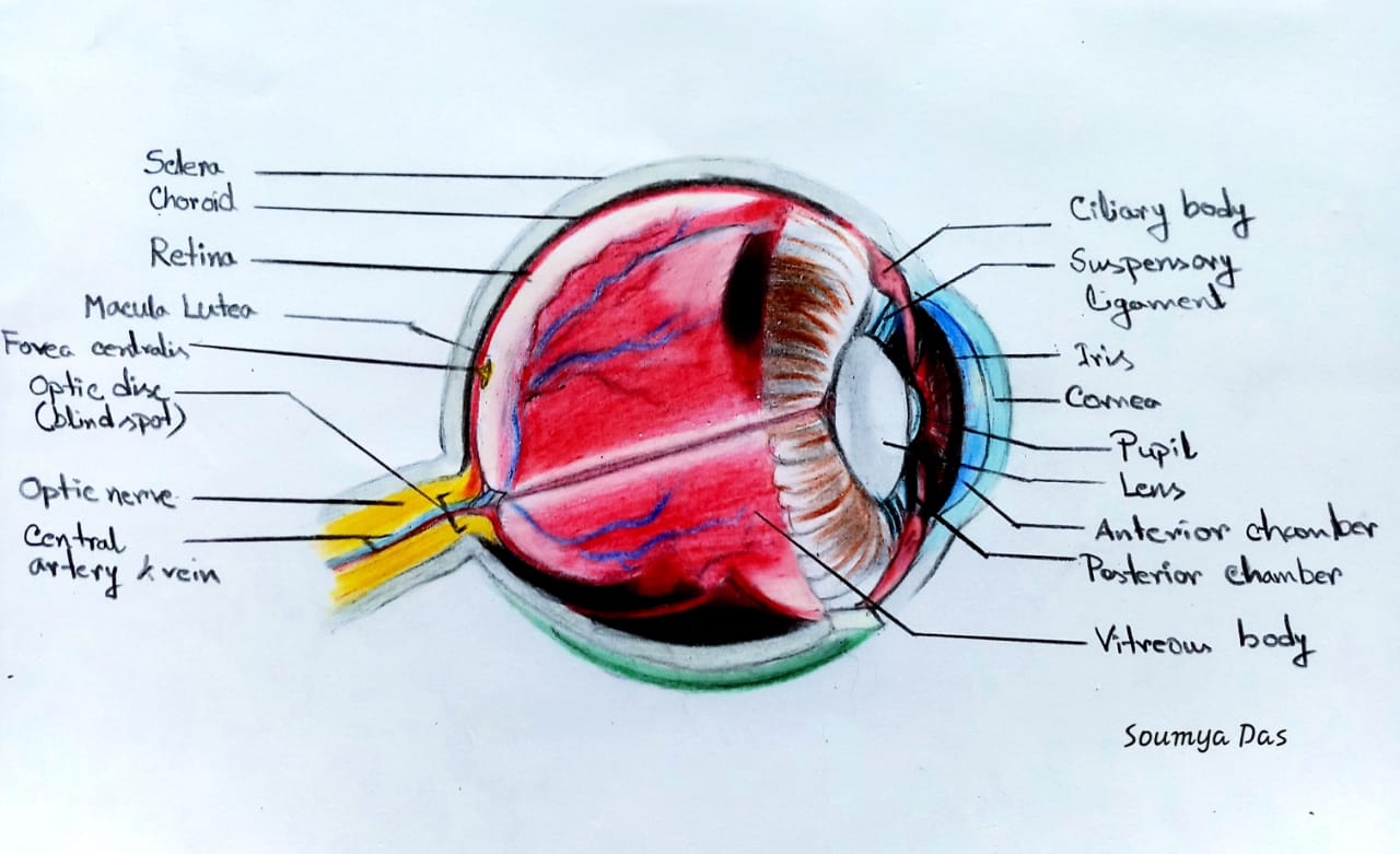
Now we can discuss the various components of the eye’s optical system.
1)The Cornea:-
The cornea is a highly transparent structure of meniscus form, approximately 12mm in diameter and slightly smaller vertically than horizontally. The center thickness is generally between 0.5 and 0.6 mm.
A thin layer of lacrimal fluid normally covers the anterior surface, but it is too thin to affect the power appreciably.
To a first approximation, both surfaces may be regarded as spherical ,the radius of curvature having values approximately +7.7 mm(anterior) and +6.8 mm(posterior).
The refractive index of the corneal substance may be taken as 1.376 and that of the aqueous humour,in contact with the back surface of the cornea, as 1.336.The power of the cornea as a whole is,therefore, about +43 D,over two-thirds of the total power of the eye.
The light entering the eye is refracted markedly at anterior corneal surface for two reasons:first because of its curvature and secondly, bacause of the big difference in refractive indices of air(1) and cornea(1.37).When the eyes are unprotected under water, the anterior surface of the cornea has its power power greatly reduced,the retinal image then becoming inordinately blurred.
The aspherical shape of cornea’s anterior surface is responsible for a baseline astigmatism of 0.25-0.5 D in almost every human eye.A changing corneal surface’s radius of curvature with distance from the centre of the pupil to the pupillary margin is responsible for a spherical aberration of 0.21-1.62 D in a normal human eye for a pupil of 4 mm in diameter.
2) The Anterior Chamber:-
From an optical point of view, the depth of the anterior chamber is important in as much as it affects the total power of the eye’s optical system.If all other elements remain unchanged, a reduction of 1 mm in the depth of the anterior chamber would increase the eye’s total power by about 1.4 D.The reverse effect would result from a shift in the opposite direction.
3)The Pupil:-
The amount of light admitted to the eye is regulated by the pupil, an approximately circular opening in the iris.The size of the blur circle on the retina generally increases with an increase in the size of the subject’s pupil, particularly one who is ametropic.With small pupil sizes,the depth of focus of the eye increases and the objects remain in adequate focus even inside the actual near point of the eye.Therefore, an artificially close near point of accommodation is measured.
4)The crystalline lens:-
The crystalline lens serves the double purpose of supplying the balance of the eye’s refractive power and providing a mechanism for focusing at different distances.This is called accommodation.
Both anatomically and optically,the lens is a highly complex structure , composed of layers of fibres placed down in an essential radial patern that is regular enough to allow a symmetrical diffraction halo to be formed.The lens continues to grow in bulk throughout life by the formation of fresh layers of fibres on the exterior.As part of the normal process of aging,it is capable to various changes impairing its flexibility and transparency.Its centre thickness is thereby increased,while the radius of curvature may become longer.
The lens has a diameter of approximately 9mm and it is biconvex in form,the radius of its anterior surface being about 1.7 times that of its posterior surface.When the lens is in its unaccommodated state,the centre thickness has traditionally been taken as 3.6mm,a figure appropriate for a young adult.As accommodation is brought into play,both surfaces but especially the anterior, assume a more steeply curved form.The central thickness thus increase and the vertex of the anterior surface moves forward, redusing the depth of the anterior diameter.
Because of its onion like structure and the compression exerted on the innermost layers,the crystalline lens is far from being optically homogeneous.It is possible to distinguish a central biconvex portion called the cortex.In the centre of the nucleus ,the refractive index reaches its maximum value between 1.40 and 1.41 but diminishes from the centre outwards , being about 1.385 near the poles and about 1.375 near the equator,the mean value being about 1.39.The entire lens has a refractive power higher than these figures indicate and would correspond to a uniform index of 1.42,if the crystalline lens was homogeneous.The total dioptric power of the crystalline lens in situ in relaxed state varies from 16 to 20D.
The assumption that the lens surfaces are spherical is for convenience only.Careful observation reveals a marked degree of peripheral flattering, especially of the anterior surface in its accommodation state.The peripheral flattering of the cornea,the eye’s spherical aberration is kept within reasonable limits.The comparatively greater refractive strength of the nucleus of the lens also diminishes the optical errors of spherical and chromatic aberration.
5)The vitreous:- The back surface of the crystalline lens is in contact with the vitreous humour,a transparent gel that fills the posterior segment of the globe.It has very nearly the same chemical composition as the aqueous and its refractive index may be taken as the same i.e. 1.336.
6)The retina:- From the optical point of view,the retina could be described as the screen on which the image is formed.It can be regarded as part of a concave spherical surface with a radius of curvature approximately -12mm.
In cameras and optical instruments usually it is convenient to have images formed on plane surfaces,but the curvature of the retina has two positive advantages.At first,the images formed by optical systems tend to have curved surfaces.The curvature of the retina is of the retina order from this point of view.Secondly,the steeply curved retina is able to cover a much wider field of view than would otherwise be possible.
Other retinal factors important to image formation:-
A) Photoreceptors (cones and rods) can be considered the pixel elements comprising a retinal image.It is the finite size of these Photoreceptors that ultimately determine the eye’s ability to resolve fine details.
B) Configuration of foveal pit and the tightly packed cones in the foveal region contribute to finest resolution of retinal image in the area of retina.
C) Orientation of retinal cones is such that they function as light pipes or fibre optic which is directed towards the second nodal point of the eye.
D) Yellow macular pigment may be considered to act as a blue filter that limits chromatic aberration and also absorbs scattered light,which is predominantly of shorter wavelength.


