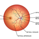
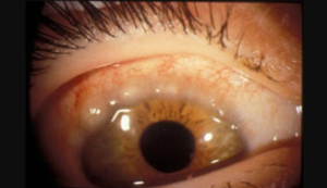
The fundus is the most important part of the eye. It is situated interior surface of the eye . It is made up of the retina, macula, optic disc , fovea and blood vessels . Retina looks like purplish –red due to rod and underlying vascular choroid.
Optic disc :- Optic disc also known as optic nerve head , it is look like pinkish colour . It is well defined vertically oval area with average dimension of 1.76mm horizontally and 1.88 mm vertically . It is placed 3.4 mm nasal to the fovea. At the optic disc , all the retinal layers terminate except the nerve fibres layer, which pass through the lamina cribrosa to run into the optic nerve. The optic disc produces an absolute scotoma in the visual field called as physiological blind spot due to absence of photoreceptors cells.
Macula lutea :- Macula lutea also known as yellow spot. In this area more darker than the surrounding fundus . It is situated at the posterior pole , is situated temporal to the optic disc. It is about 5.5 mm in diameter . Fovea centralis is the central depressed part of the macula.
Techniques of fundus examination :-
Fundus examination is essential to diagnose the diseases of the vitreous , optic nerve head , retina and choroid .
For fundus should be dilated with 5% phenylephrine and 1% tropicamide eye drops.
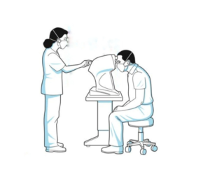
Fundus examination can be examined by Ophthalmoscopy , slit lamp biomicroscopy .
Ophthalmoscopy :- Ophthalmoscope is a device which is used to examine your eyes interior structure , including the retina and the procedure is called ophthalmoscopy . Ophthalmoscopy was first invented by Babbage in 1848 and it is modified by Von Helmholtz in 1850. Three types of ophthalmoscopy was vogue introduced such as ;-
Distant direct Ophthalmoscopy , Direct Ophthalmoscopy and Indirect Ophthalmoscopy .
Direct Ophthalmoscopy :- It is the method which are commonly used in Ophthalmoscopy . The direct ophthalmoscopy works on the basics principle of glass plate ophthalmoscope which was introduced by Von Helmholtz. In this Ophthalmoscopy a convergent beam of light is reflected into the patients pupil. The emergent rays from any point on the patient’s fundus reach the observer retina through the viewing hole in the ophthalmoscope . The emergent rays from the patient’s eye are parallel and brought to focus on the retina of the emmetropic observer when accommodation is relaxed . However , if the patient and the observer are ametropic , a correcting lens must be interposed .
In direct Ophthalmoscopy , the image is erect , virtual and about 15 times more magnified in emmetropes .
Direct Ophthalmoscopy should be performed in a semi dark room , patient seated and look straight ahead , while observer standing or seated slightly over to the side of the eye to be examined . Patient right can be examined by the observer right eye .
Distant direct ophthalmoscopy :- It should be performed routinely before the direct ophthalmoscopy , as it gives a lot of useful information. It can be performed with the help of a self illuminated ophthalmoscope . The light is thrown into patient’s eye sitting in a semi darkroom , from a distance of 20 -25 cm and the features of the red glow in the pupillary area are noted.
- To diagnose opacities in the refractive media :- Any opacities in the refractive media is seen as a black shadow in the red glow. The exact location of the opacity can be determined by observing the parallactic displacement.
- A small hole and a mole on the iris appear as a black spot on oblique illumination . On distant direct ophthalmoscopy , the mole looks black but a red reflex is seen through the hole in the iris .
- A greyish reflex seen on distant direct ophthalmoscopy indicates either a detached retina or a tumour arising from the fundus.
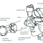
Indirect Ophthalmoscopy :- indirect Ophthalmoscopy was introduced by Nagel in 1864. The principle of indirect ophthalmoscopy is to make the eye highly myopic by placing a strong convex lens in front of patient’s eye so that the emergent from an area of the fundus are brought to focus as a real , inverted and magnified image .
In indirect ophthalmoscopy patient is made to lie in the supine position. The examiner throws the light into patient’s eye from an arm’s distance . the examiner looks the different movement of the beam of light .
Slit lamp biomicroscopy :-
Biomicroscopy examination of the fundus can be examined by three type of lens
- Indirect fundus biomicroscopy can be done by +78D or +90D lens. In this lens image is formed inverted , real and magnified.
- Hruby lens biomicroscopy :- Hruby lens is a plano concave lens with 58.6D lens, in this lens image is formed with minified.
- Contact lens biomicroscopy :- it can be performed by two type of lens such as posterior fundus contact lens and Goldmans’s three – mirror contact lens.

