
What is the meaning of Refractometry ?
It is the estimation of refractive error with a machine called refractometer or optometer.
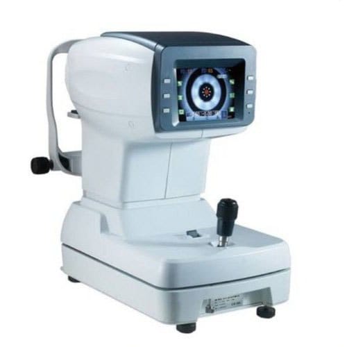
Autorefractometry
- An autorefractor is an alternative method of finding out of error of refraction by the use of an optical equipment called refractometer or optometer.
- The process is quick and painless for the patient, and the data ensures a baseline to determine the correct eyeglass and contact lens prescription.
OPTICAL PRINCIPLES :-
- Scheiner Principle – 1619
Scheiner{1619} observed that refractive error of eye can be determined by using double pinhole apertures.
Parallel rays of light entering the eye from a distant object,are limited to two small bundles when double pinhole apertures are placed in front of the pupil.
Myopia bundles cross each other before reaching the retina two small spots of lights are seen.
Hypermetropica rays are intercepted by the retina before they meet àagain two small spots are seen.
These two points of light can be coalesced to a single point by moving the double pinhole to the far point of eye.
So from far point of eye,refractive error of eye can be found.
- Myopia
- Hyper-metropia
- Optometer principle – 1759
Porterfield{1759}à coined term optometer.
Use a single converging lens placed at its focal length from the eye instead of interchangeable trial lenses.
Light from the target on far side of lens enters the eye with vergence of zero/minus/plus, depending on the position of target.
Vergence of light in the focal plane of optometer lens is linearly related to the displacement of target.
A scale with equal spacing can thus be made which would show the no. of dioptres of correction. ( optometer principle)
COMMERCIALLY AVAILABLE OBJECTIVE AUTOREFRACTOMETR:-
- The Scheiner principle
- The optometric principle ( retinoscopic principle)
- The best -focus principle
- The knife – edge principle
- The ray – deflection principle
- The image size principle
- How Does an Autorefractor Work?
The autorefractor shines light into the eye, measuring how it changes as it bounces off the back of the eye (the retina). Then, the patient is shown an image that moves in and out of focus. As reflections are made, several measurements determine when the eye is properly focused. These final figures determine the level of vision correction needed. Unlike many conventional refraction diagnostic devices, some autorefractors utilizes a unique wavefront technology algorithm based on Hartmann-Shack wavefront sensor, which analyzes many focal spots of a light wavefront. It can measure not just the basic refraction error of a patient, but to obtain a spatially resolved refraction map of the wavefront.
Procedures of using autorefractor
- For autorefractor testing, ask your patient to be seated on a chair in the diagnostic procedure room.
- Your patient’s head will then be positioned so that their chin is resting on the chin rest and their forehead is resting on the forehead rest of the autorefractor.
- Then ask your patient to look on the target through right eye.
- Now you have to move the joystick to push the device left/right or up/down or front/back so that the image of the patient’s eye that you are viewing in the monitor is focused.
- Once the image is on focus, you must position the square on the center of the monitor at the pupil reflex.
- Once the square is positioned at the corneal reflex and the patient’s eye image is on focus you have to press the capture button on the joystick for three times
- When the capture button is pressed, the machine bounces off infra-red rays from the retina (innermost light sensitive layer of the eye) back to device and takes a series of measurements to determine the patient’s refractive power. It will show three different reading of the patient’s refractive error and gives the average reading as the final reading for the refractive error.
- Finally the device will print out the final prescription (average reading from the three readings) of the patient’s refractive error.
- Repeat the above procedures for left eye.


