
What is DCR?
When the tear duct is obstructed, tears cannot drain properly from the eye then a surgical procedure is performed to treat a blocked tear duct.
There are two procedure to treat this blocked duct
1.DCT ( Dacryocystectomy)
2. DCR (dacryocystorhinostomy)
Why is DCR performed?
The Tear duct, also known as nasolacrimal duct, is responsible for draining tears from the eyes into the nasal cavity. When this duct become blocked due to some reason, leading to symptoms such as
1. Watering from the eye
2..Discharge from the eyes
3. Eye infection
4. Irritation and discomfort
5. Sometimes Excessive pain
6.Swealling in the nasolacrimal duct
DCR or DCT is generally suggested by an eye care professional when the other’s primary treatment doesn’t work or fails to give resolution to the patient from this problem, such as Antibiotic Medicine, Tear duct probing etc.
During DCR a new pathway is created for passing the tears and drain directly in the nasal cavity .
Why do tears duct become blocked?
There are various reasons for blocking in the Nasolacrimal Duct
1. Congenital Anomalies: Some are born with abnormalities that can cause narrowing or obstruction of the tears Ducts.
2. Aging: As people age the teats duct system becomes less efficient due to changes in tissue structure, leading to blockages.
3. Inflammation or Infection: Inflammation or infection of the tear ducts can lead to swelling and blockage.
4. Trauma : Any injury on the face or eye region can lead to blockage in the Tears drainage system.
5. Sinus or Nasal Condition: This can affect the nasal passages or sinuses such as chronic sinusitis or nasal polyps can cause pressures on the tears ducts.
6. Medications: Certain Medications, such as antihistamines or decongestants can dry out the eyes and lead to thickening of tear fluid. Sometimes chronic dry eye syndrome can lead to inflammation and scarring of the tears ducts.
7. Idiopathic: In some cases, the exact cause of tear duct blockage may not be identified and it may occur without an apparent underlying reason.
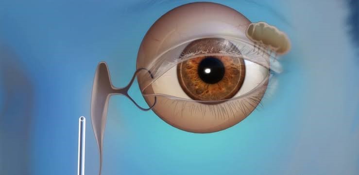
How tears drain?
Tears automatically drain out from the conjunctival sac to capillary forces of lacrimal puncta that present on the inside corners of our upper and lower eyelids, which suck up the liquid into the lacrimal canaliculi. Canaliculi move tears to a sac to a reservoir on the side of the nose known as lacrimal sac. This whole procedure is stimulated by orbicularis oculi muscle contraction.
How is the DCR operation performed?
1. Preparation: The patient is positioned comfortably on the operating table, usually under local or general anesthesia, depending on the surgeon’s preference and the patient’s medical condition.
2. Access: The surgeon typically approaches the lacrimal sac through the nasal cavity. This can be done either externally (external DCR) or endoscopically (endoscopic DCR). The surgeon makes a small incision in the skin near the inner corner of the eye for external DCR, or in the nasal cavity for endoscopic DCR.
3. Exposure: Using specialized instruments and techniques, the surgeon carefully exposes the lacrimal sac and the area where the new drainage pathway will be created.
4. Creation of Osteotomy: In both external and endoscopic DCR, the surgeon creates an opening (osteotomy) in the bone of the nasal cavity, typically near the lacrimal sac. This opening allows access to the lacrimal sac and creates a pathway for tears to drain into the nasal cavity.
5. Removal of Obstructions: Any obstructions or scar tissue blocking the tear drainage pathway are removed to ensure proper tear flow.
6. Mucosal Flap: In endoscopic DCR, a mucosal flap may be created from the lining of the nasal cavity to cover the osteotomy site and help prevent scarring.
7. Placement of Stent or Tube (Optional): In some cases, a silicone stent or tube may be inserted into the newly created drainage pathway to keep it open during the healing process. This helps maintain the patency of the pathway and prevents it from closing prematurely.
8. Closure: The incisions in the skin or nasal cavity are closed with sutures, and a nasal pack may be placed to minimize bleeding and provide support to the newly created drainage pathway.
9. Postoperative Care: The patient is monitored closely in the recovery room and given instructions for postoperative care, which may include nasal irrigation, antibiotic eye drops, and pain management.
10. Follow-up: Follow-up appointments are scheduled to monitor the healing process and assess the success of the surgery. The silicone stent or tube, if used, may be removed several weeks after surgery.
Actual goal of a DCR operation is to create a new pathway for drainage of the tears properly and relieve them from the symptoms that are associated with nasolacrimal duct obstruction.
The procedure of DCR surgery may vary depending on the Surgeon’s technique and anatomy of nasolacrimal duct and patient medical history.
If you’re a non medical person and you think you are experiencing this issue, it is advisable to visit an eye care professional for a comprehensive consultation and examination, it can provide detailed guidance and recommendations.


