
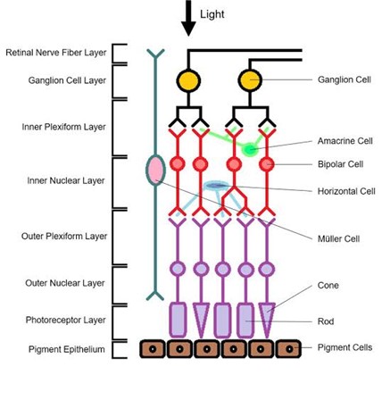
Layers of the retina, which is a complex and essential part of our visual system. Within the retina, there are ten distinct layers, each contributing to our ability to perceive light and form visual images. Here’s a comprehensive overview of these layers:
1. Retinal Pigment Epithelium (RPE):
- The outermost layer of the retina, adjacent to the choroid.
- Composed of pigmented cells that absorb excess light and prevent reflection
back into the neurosensory layer. - Vital for maintaining the health of photoreceptor cells.
2. Photoreceptor Layer:
Contains two types of photoreceptor cells:
- Rods: Sensitive to low light conditions and provide monochromatic vision.
- Cones: Function in well-lit conditions and allow us to perceive colors and details.
- Rods and cones contain specialized pigments that absorb light.
3. Outer Nuclear Layer (ONL):
- Houses the cell bodies of photoreceptor cells.
- The density of nuclei varies, with cones being more
- Concentrated in the fovea and rods distributed more peripherally.
4 . Outer Plexiform Layer (OPL):
- Synaptic connections between photoreceptor cells and bipolar cells.
- Bipolar cells transmit signals from photoreceptors to ganglion cells.
5. Inner Nuclear Layer (INL):
- Contains the cell bodies of bipolar cells, horizontal cells, and amacrine cells.
- Bipolar cells relay signals from photoreceptors to ganglion cells.
- Horizontal and amacrine cells modulate communication between different layers.
6. Inner Plexiform Layer (IPL):
- Synaptic connections between bipolar cells and ganglion cells.
- Ganglion cells are the output neurons of the retina, sending signals to the brain via the optic nerve.
7. Ganglion Cell Layer (GCL):
- Houses the cell bodies of ganglion cells.
- Ganglion cells integrate visual information from bipolar cells and transmit it to the brain.
- Some ganglion cells are specialized for detecting motion, while others contribute to colour vision.
8. Nerve Fiber Layer (NFL):
- Contains the axons of ganglion cells.
- These axons converge to form the optic nerve.
- which carries visual information to the brain.
9. Inner Limiting Membrane (ILM):
- The innermost layer of the retina, separating it from the vitreous humor.
- Composed of specialized glial cells (Müller cells) and acts as a barrier.
10. Macula and Fovea:
- The macula is a small, specialized area in the central retina.
- Within the macula, the fovea is the center of highest visual acuity.
- The fovea contains a high density of cones and is responsible for sharp, detailed vision.
■ Anatomy of the Retina:
- The retina is a thin layer of neural tissue that lines the entire back wall of the eye. It’s the only extension of the brain visible from outside the body during a retinal exam.
- Key components of the retina include:
Photoreceptors: These are specialized light- sensitive cells responsible for
turning light into signals that are sent to the brain. There are two main types:
Rod cells: Extremely sensitive to light, they help us see in dim light conditions.
Cone cells: Provide color vision and help us see fine details. There are three types of cone cells: red, green, and blue.
Macula: Located at the retina’s center, the macula contains a high concentration of cone cells. It helps us see details in the center of our visual field.
Fovea: The very center of the macula, where vision is sharpest due to the
abundance of cone cells.
Peripheral Retina: Outside the macula, responsible for peripheral vision and night vision. Rod cells are more abundant in this area.
■ Functions of the Retina:
The retina converts incoming light into visible images. It acts as a bridge
between the eye and the brain.
When light rays enter the eye through the pupil, the retina creates electrical
signals. These signals travel through the optic nerve to the brain's visual
cortex, where they are processed into recognizable images.
Essentially, the retina turns light energy into neural signals that the brain
interprets as images.


