Diplopia usually results from an acquired misalignment of the visual axes that causes an image to fall on the fovea of one eye and simultaneously on a non foveal point in the other eye. The object that falls on these non corresponding points must be outside Panum’s area to be seen double.
In diplopia, the same object is seen as having two different locations in subjective space; the foveal image is always clearer than the non foveal image.
UNDERSTANDING THE CAUSES OF DIPLOPIA
1. Confusion: It occurs due to formation of image of two different objects on the corresponding points of two retinae following misalignment of the visual axes of two eyes .
2. Ocular deviation: In paralytic strabismus, the primary ocular deviation is in comitant and differs from the secondary deviation. However, with the passage of time there occurs spread of comitance.
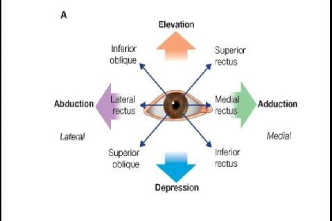
Primary deviation: It is deviation of the affected eye, when the unaffected eye is used for fixation and is away from the action of paralyzed muscle, e.g. if lateral rectus is paralyzed, the eye is
convergent.
Onset of paralytic ocular deviation may be of sudden as seen in trauma and vascular occlusions; or gradual as seen in tumor’s and multiple sclerosis.
Secondary deviation: It refers to deviation of the unaffected eye seen under cover, when the patient is made to fix with the affected eye. In a recently acquired ocular palsy, the secondary deviation is much greater than the primary deviation. This is due to the fact that the strong impulse of innervation required to enable the eye with paralysed muscle to fix is also transmitted to the yoke muscle of the sound eye resulting in a greater amount of deviation. This is based on Herring’s law of equal innervation of diag yoke muscles.
3. Ocular movements: Restriction of ocular Movements occurs towards the direction of action of the paralyzed muscle/muscles. When the paralysis is of recent onset, a careful study of duction and version movements will make the diagnosis on the basis of incomplete movements in the field of action of the paralyzed muscle. However, in long-standing cases, development of muscle sequelae such as contracture of the direct antagonist muscle and secondary inhibition palsy of the contralateral antagonist muscle, present difficulties in the identification of the paralyzed muscle. Under these circumstances, other tests like head-tilt test and Hess screen charting, etc. may be helpful in diagnosing the paralyzed
muscle.
4. Past pointing. Past pointing also described as false projection or orientation occurs due to increased innervational impulse conveyed to the paralyzed muscle during movement in the direction of action of paralyzed muscle.
Treatment of double vision
A. Diplopia exercise the aim is to make the patient aware of physiological diplopia in
heterophoria and inter- mittent tropia and of diplopia in heterotropia.
1. Awareness of physiological diplopia: Awareness of physiological diplopia is an effective means of treating suppression in patients with phorias, intermittent tropias and phorias recently converted from
tropias by surgery and/or orthoptics. Patients should be taught to experience both homonymous and heteronymous physiological diplopia . This will stimulate both the nasal and temporal elements of
the retina and thus will help the patient to overcome suppression over the entire retina.
I. Awareness of homonymous (uncrossed) diplopia. Patient is asked to make note of light situated about 6 meeters from him/her while fixating a light held 33 cm directly in front of his/her nose. While doing so, by practice, patient will learn to see one light at near (33 cm) and two lights at distance (6
meters) simultaneously.
II. Awareness of heteronymous (crossed) diplopia. Patient is asked to make a note of light held 33 cm in front of his/her nose while fixating a light situated 6 meters away. While doing so, by practice, patient will learn to see one light at distance and two lights at near simultaneously.
III. Exercises utilizing physiological diplopia
Framing: Framing is a good home exercise based on physiological diplopia. Patient is asked to make note of a pencil held at 15 cm in front of his/her nose while fixating at a distant object in the room. Patient should see two pencils and one distant object (awareness of heteronymous diplopia) . Now the patient is asked to move the pencil away from his/her nose. During this procedure, patient will note that two pencils are moving closer to each other. Similarly, when the pencil is moved towards the nose, its two images will be seen moving farther apart from each other. Once the patient learns this, he/she is asked to fix at different distant objects in the room and move the pencil backward or forward so that the distant object is framed between the two pencils, i.e. the object is centred with pencils touching its each side .
Bar reading. Bar reading exercise is also based on the principle of awareness of crossed physiological diplopia. Patient is asked to hold bar (thumb, finger, pencil or any other bar) about 5-6 cm from his/her nose and asked to read a print kept at about 33 cm from his/her eyes . As the patient reads the print binocularly, he/she perceives the bar in crossed diplopia, each image of the bar hiding a portion of the print from one eye, but not the other, so that patient can read the print normally. Such a practice of maintaining the correct position of his/her eyes despite the obstacle will strengthen the binocular vision of the patient.
2. Diplopia exercises with coloured filters: These exercises are based on the fact that a patient with strabismus suppresses similar images in contrast to normal person who tends to suppress dissimilar images. In this exercise, patient is asked to fixate a white light and a red filter is placed in front of one eye and a green filter in front of the other eye. The dissimilar images produced by the filters are sufficient to overcome suppression in most instances and produce DIPLOPIA.
3. Diplopia exercise with prisms: In this orthoptic exercise, a vertical prism (base up or down) sufficient enough to displace the image outside the suppression scotoma is placed in front of one eye. Consequently, the patient will have diplopia. Gradually, the prism power is reduced until finally the patient has diplopia without any vertical displacement of one image. Once the patient learns to perceive diplopia without prism, the fixation light is then replaced by a less intense stimulus. In this way, patient should be taught diplopia
during both near and distance fixations.
(B) Exercises with use of red filter
1. To treat suppression at near, a red filter is placed over the dominant eye and patient (depending upon his/her age and interest) is asked to do any of the following exercises:
• Colour, trace or draw with a red pencil matching the red filter.
• Little girls may follow red design drawn on a white cloth with a needle and a light red or pink thread.
• Children can draw lines with a red pencil using the number-to-number games in their coloring books.
• Children can also select red beads for a necklace
• The boys can play ping-pong with a red ball.
2. To treat suppression at distance, patient may be asked to watch colour television with the red filter over the dominant eye.
C. Exercises with major amblyoscope
1. Macular massage. This exercise stimulates the retina of deviated eye. It is accomplished by moving the visual target on the major amblyo- scope back and forth across the suppression scotoma (macular massage) as below:
The major amblyoscope tubes having slides of paramacular simultaneous perception are locked at the patient’s objective angle. While he/ she looks constantly straight ahead, the tubes are moved rapidly from side to side over a large arc. Since they are at the objective angle, the images moving over the retina will always stimulate normally corresponding areas. In the periphery, they will be superimposed.
2. Crossing technique. A pair of paramacular simultaneous perception slides is put in the tubes of major amblyoscope. Patient is asked to fixate the target viewed by the dominant eye, while the target in front of the suppressed eye is moved in from the periphery of the field towards the suppression scotoma. It will disappear, when it reaches the suppressed area and reappear on the other side after the entire scotoma has been crossed. This back and forth movement of the target across the suppression area is continued until this area has decreased to such an extent that the patient can simultaneously perceive both targets and can superimpose the two images. In normal correspondence,
peripheral size targets may be used first followed by macular and foveal size targets.
3. Chasing technique. It is a technique of subjective exercise using the smallest simultaneous perception slides that the patient can superimpose. The two arms of the major amblyoscope are loosened and the patient is asked to hold the handle of the tube in front of the suppressed eye. The examiner moves the picture tube that is in front of the fixating eye in a random position. The patient is asked to chase it and superimpose the two pictures by moving the other tube. This chasing technique is exercised repeatedly. As the patient's performance improves, increasingly smaller pictures
are used until he/she can superimpose the foveal slides.
D.Exercises with chireoscope
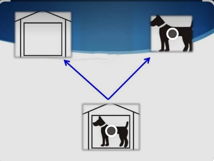
The cheiroscope is an instrument for anti-suppression exercises. It consists of a working base, a picture carrier to one side, a headrest containing a pair of +7.0D spherical lenses, and an obliquely placed septum extending from the center point between the lenses to the base of the picture carrier. A plane mirror is attached to the septum on the side where the picture is located. The distance between lenses and the working base is 14 cm, which is the focal length of each lens and the eyes are
consequently focused for distance.
Procedure: The picture to be copied is placed in the picture carrier and a sheet of paper on the base of the instrument. The patient should look with his/her fixating eye into the mirror and with the suppressing one on the paper. He/she is instructed to trace the picture (which has black outlines) on the paper using a pencil (a red- colored pencil is very helpful, especially in the
beginning). Steady fixation of the picture should be stressed to prevent rapid alternation, which can be suspected, if the patient’s drawing is either smaller or larger than the picture or if parts of the picture are missing.
E. Exercises with Tibb’s binocular trainer The Tibb’s binocular trainer is a haploscopic instrument designed for home use.
Procedure : one wing board is placed so that it rest along the table at a slight
angle to it . The vertical wing board is to the right side, when the right
eye is the fixating one; it is to the left side, when the left eye is fixating.
The patient places the bridge of his/her nose against the curved part of
the septum so that his/her visual axis is perpendicular to the table.
His/her head should not be tilted.


