
Examination of ocular movements involves the assessment smooth pursuit movements followed by saccades.
Types of Eye Movements
Ductions
These are monocular movements from one position to another. They consists of followings
- Adduction ( Medial Movement) – Occurs due to action of medial rectus.
- Abduction ( Lateral Movement) – Occurs due to action of lateral rectus.
- Supraduction ( Elevation) – Occurs due to action of superior rectus assisted by inferior oblique.
- Infraduction ( Depression) – Occurs due to action of inferior rectus assisted by superior oblique.
- Incycloduction ( Intorsion) – Occurs due to action of superior oblique.
- Excycloduction ( Extorsion) – Occurs due to action of inferior oblique.
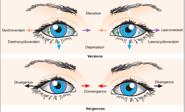
Versions
These are binocular and conjugate movements ( in the same direction so the angle b/w eyes remains constant).
These are followings
- Dextroversion ( Right Gaze) – Occurs due to action of right lateral rectus and left medial rectus.
- Levoversion ( Left Gaze) – Occurs due to action of left lateral rectus and right medial rectus.
- Dextroelevation ( Gaze up and right) – Occurs due to action of right superior rectus and left inferior oblique.
- Levoelevation ( Gaze up and left) – Occurs due to action of left superior rectus and right inferior oblique.
- Dextrodepression ( Gaze down and right) – Occurs due to action of right inferior rectus and left superior oblique.
- Levodepression ( Gaze down and left) – occurs due to action of left inferior rectus and right superior oblique.
- Dextrocycloversion: Occurs due to extorters of right eye i.e inferior rectus and inferior oblique and intorters of left eye, namely, superior rectus and superior oblique.
- Levocycloversion: Occurs due to action of extorters of left eye and intorters of right eye.
Vergences
These are binocular and disjugate movements ( in opposite direction so the angle b/w the eyes change).
- Convergence is inward or medial turning of eyes due to action of both medial recti. It may be voluntary or reflex.
- Divergence is outward or lateral turning of eyes that is produced due to action of both lateral recti.
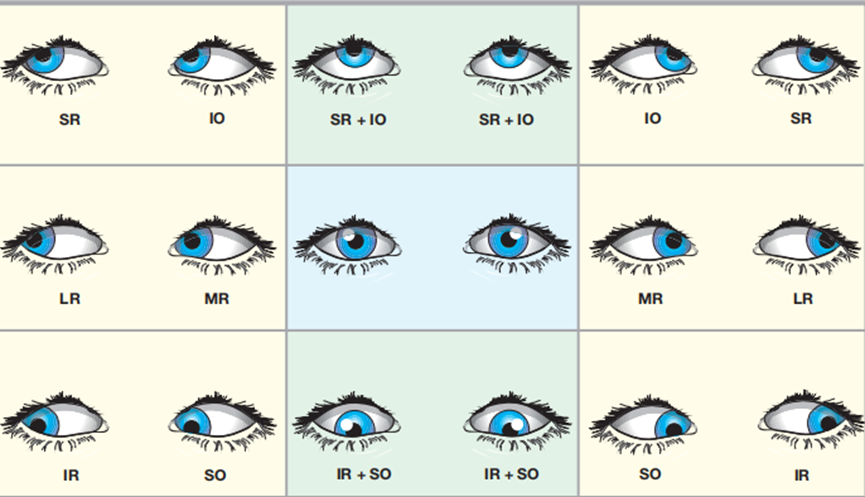
Positions of Gaze
Six Cardinal Positions of gaze are identified as which muscle is responsible for moving the eye into a particular position
- Dextroversion ( Right L.R + left M.R).
- Levoversion ( Left L.R + Right M.R).
- Dextroelevation ( Right S.R + Left I.O).
- Levoelevation ( Left S.R + Right I.O).
- Dextrodepression ( Right I.R + Left S.O).
- Levodepression ( Left I.R + Right S.O).
Nine diagnostic Positions of gaze are those in which deviations are measured.
They consist of six cardinal positions, primary position, elevation and depression.
Primary Position of eye is when eye is looking straight ahead.
Secondary Position of the eye is in adducted, abducted, elevated and depressed positions.
Tertiary Position of the eye is in oblique position.
Laws of Ocular motility
Sherrington law:
It states that increased contraction of a muscle is accompanied by decreased contraction of its antagonist.
It means that when medial rectus contracts, lateral rectus automatically relaxes and vice versa.
It applies to both versions and vergences.
Hering law:
It states that during any conjugate eye movement, equal and simultaneous innervation flows from brain to the yolk muscles.
For example, During right gaze, the right lateral rectus and left medial rectus receive equal and simultaneous innervation.
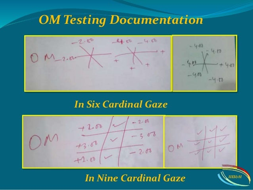
Procedure
The purpose of ocular motility evaluation is detection of palsy checking the function of extra ocular muscles and diplopia.
Patient is seated and movements of eyes are checked in all positions of gaze by asking the patient to follow a torch light. By use of torch light, corneal reflections can also be checked to help in assessment.
A quick Cover test is performed in every position of gaze to detect deviation and patient is questioned regarding diplopia.
Vertical and horizontal saccadic movements are checked by asking the patient to look rapidly b/w widely separated targets. In non cooperative patients, versions are elicited by noise and doll’s head manoeuvre.
In case of palsy e.g in left lateral rectus palsy, eye will not move outward and will deviate inward. It shows over action of medial rectus and under action of lateral rectus. Any attempt to move the eye to left will cause diplopia.
Over action of a muscle is denoted by + sign and under action is denoted by – minus sign.


