
In the realm of clinical optometry and vision science, dynamic retinoscopy stands as a time- tested and indispensable technique for evaluating functional vision performance. Particularly when it comes to understanding accommodative disorders, this method offers direct, real- time insight into how the eyes are truly functioning in natural, near-vision tasks. As our understanding of visual function becomes more holistic, dynamic retinoscopy has become central to diagnosing, differentiating, and managing a variety of accommodative anomalies.
Understanding Dynamic Retinoscopy:
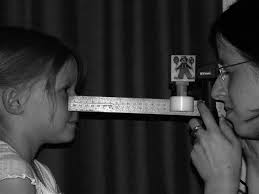
Dynamic retinoscopy differs from its static counterpart by assessing the eye's focusing ability under natural, near-working conditions. While static retinoscopy is conducted with the patient fixating at a distance (usually 6 meters or more), dynamic retinoscopy evaluates the accommodative response while the patient fixates on a near target, typically at 40 cm. This method provides invaluable data on how accurately and efficiently the eyes accommodate in real-time situations, such as reading, writing, or using digital screens. By observing the reflex of the retinoscope beam as it interacts with the patient’s pupil, the clinician can judge whether the patient is over-accommodating, under-accommodating, or showing signs of fatigue or instability in their accommodative system. The beauty of dynamic retinoscopy lies in its objectivity: the test does not rely on the patient’s subjective responses, making it especially useful in pediatric, non-verbal, or neurologically impaired populations.
Accommodative Disorders: An Overview
Accommodation is the eye’s ability to change its optical power to maintain a clear image (focus) as objects move closer or further away. This process involves a dynamic interplay between the ciliary muscles, lens elasticity, and neurological control. When any component of this system fails to function correctly, it leads to accommodative disorders. These can be broadly classified into:
1. Accommodative Insufficiency (AI): The eye fails to stimulate adequate accommodation for near tasks. Common in children and young adults, it manifests as blurry near vision, difficulty reading, and eye strain.
2. Accommodative Excess (AE): Also known as accommodative spasm, this condition features an overactive accommodative response, making it difficult for the eye to relax focus for distance vision.
3. Accommodative Infacility (AIF): The eye struggles to shift focus efficiently between near and far objects, leading to delayed visual clarity, particularly in dynamic environments.
These conditions often overlap in symptoms: headaches, visual discomfort, reduced reading endurance, and difficulty with academic or office tasks. Without precise diagnostic tools, these disorders can go undiagnosed or be mistaken for learning disabilities, attention deficits, or behavioral problems.
Techniques of Dynamic Retinoscopy
There are several standardized methods of dynamic retinoscopy, each suited to different clinical goals and patient profiles:
1. Monocular Estimation Method (MEM):
This is the most widely used technique. The patient views a near card monocularly while the clinician briefly introduces lenses in front of the eye to neutralize the reflex. A neutral reflex indicates a normal accommodative response. A "with" movement (lag) indicates under-
accommodation; an "against" movement (lead) suggests over-accommodation.
2. Nott Retinoscopy:
Instead of using lenses, the examiner changes their distance from the patient to identify the point of neutrality. This method is more naturalistic and can be more accurate in certain settings since it does not disrupt the patient’s accommodative state with lens changes.
3. Book Retinoscopy:
This technique is more qualitative and observational. It involves prolonged fixation on a reading passage while the examiner observes changes in the reflex. This method helps identify fluctuations in accommodation over time, often revealing fatigue or instability not
seen in brief tests.
Diagnostic Power of Dynamic Retinoscopy
The main advantage of dynamic retinoscopy is its ability to quantify the *lag or lead of accommodation:
Normal Lag: About +0.25 to +0.75 diopters at near.
Excessive Lag: Greater than +0.75 D, indicative of AI or accommodative fatigue.
Lead of Accommodation: Less than +0.25 D or negative values, may indicate AE.
Additionally, inconsistent or fluctuating reflexes can suggest issues such as accommodative
infacility or visual fatigue, often linked to prolonged digital device use or attentional issues.
Dynamic retinoscopy not only confirms the presence of accommodative dysfunction but also
helps differentiate between disorders, guiding further intervention. For example, a consistent
high lag may warrant plus lens intervention for near work, while fluctuating reflexes might
indicate the need for accommodative facility training.
Application in Different Populations
Paediatric Patients:
In children, particularly those with reading difficulties or suspected learning disorders, dynamic retinoscopy is a game-changer. Many children are misdiagnosed with attention deficits when the root cause is an unrecognized accommodative issue. Since the test does not require verbal responses, it's ideal for younger or non-verbal children.
Patients with Neurological Conditions:
Children or adults with traumatic brain injuries (TBI), cerebral palsy, or developmental delays often present with subtle but significant accommodative anomalies. MEM or Nott-retinoscopy offers objective insight into their visual functioning and helps tailor vision
therapy or prescribe optical aids.
Adults with Visual Fatigue:
In today”s digital world, accommodative fatigue and infacility are increasingly common among office workers and students. Dynamic retinoscopy helps detect early signs of visual stress that may not be apparent on subjective tests, allowing timely intervention through vision therapy or ergonomic advice.
Integrating Dynamic Retinoscopy into Clinical Practice
Despite its proven value, dynamic retinoscopy is often underutilized due to perceived complexity or time constraints. However, with practice, it becomes a quick and efficient test that can be seamlessly integrated into routine eye exams, especially for children, students, oranyone presenting with visual discomfort.
Clinicians can consider the following tips:
Keep a set of near cards and trial lenses ready at all times.
1. Use engaging fixation targets (e.g., age-appropriate reading material or cartoons).
2. Record findings systematically and correlate them with symptoms and other test results.
Dynamic retinoscopy also enhances patient education. Demonstrating accommodative
dysfunction through objective methods increases compliance with vision therapy and near
prescriptions.
Conclusion
Dynamic retinoscopy is a cornerstone technique in the evaluation of accommodative function. Its simplicity, objectivity, and diagnostic power make it invaluable for optometrists and vision therapists alike. In an era where visual demands are rapidly increasing, especially at near, understanding and managing accommodative disorders is more important than ever. By integrating dynamic retinoscopy into regular clinical workflows, eye care professionals can uncover hidden visual problems, provide targeted interventions, and ultimately improve the quality of life for their patients. In the hands of a skilled clinician, dynamic retinoscopy does more than diagnose—it reveals how a person truly sees and interacts with their world.


