
Illuminating Insight: The Diverse Functions of Fluorescence
Staining in Ocular Examination and Diagnosis
Introduction:
In the realm of ocular examination and diagnosis, fluorescence staining
emerges as a powerful tool, offering clinicians a window into the intricate
structures of the eye. Through the application of fluorescent dyes, various
aspects of ocular health can be illuminated, aiding in the detection and management of a wide array of conditions. Let’s delve into the multifaceted functions of fluorescence staining in ocular care, along with its
mechanism of action and why fluorescence is utilized.
Corneal Abrasions and Ulcers Detection:
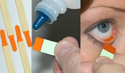
Fluorescein, a commonly used fluorescent dye, owes its illuminating
properties to its mechanism of action and the unique characteristics of
fluorescence. When applied topically  to the eye, fluorescein penetrates areas of disrupted corneal epithelium due to its low molecular weight. Upon exposure to cobalt blue light, fluorescein emits a bright green fluorescence, accentuating the areas of corneal injury. This phenomenon occurs because fluorescein molecules absorb light energy at one wavelength (blue light) and then emit it at a longer, visible wavelength (green light), resulting in the characteristic green glow that highlights corneal abnormalities.
Tear Film Evaluation:
Fluorescein and lissamine green dyes facilitate tear film evaluation through distinct mechanisms. Fluorescein, when added to the tear film, disperses evenly in the presence of a healthy lipid layer. However, disruptions in tear film stability or integrity lead to irregular fluorescein distribution, with areas of tear film breakup appearing as dark spots under cobalt blue light. Lissamine green, on the other hand, stains devitalized epithelial cells directly, aiding in the identification of areas of ocular surface damage.
Conjunctival Abnormalities Identification:
Rose bengal and lissamine green dyes serve as vital tools in visualizing conjunctival abnormalities. These dyes adhere to devitalized epithelial cells and mucous strands due to their affinity for damaged tissue. Under cobalt blue light, these areas appear as bright spots or streaks, allowing for the detection of conjunctival irregularities and the differentiation of healthy from damaged tissue.
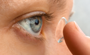
Contact Lens Fitting and Assessment:
Fluorescein staining facilitates contact lens assessment by highlighting areas of insufficient tear exchange or improper lens fit. When applied with the lens in place, fluorescein dye accumulates in regions where the lens interacts with
the ocular surface. Areas of poor fit or inadequate tear exchange appear as dark regions under cobalt blue light, guiding adjustments to optimize lens fit and ocular health.
Corneal Foreign Body Localization:
Fluorescein staining aids in the localization of corneal foreign bodies by accentuating disruptions in the corneal epithelium. When a foreign object penetrates the cornea, it creates a defect that fluorescein dye readily fills. Under cobalt blue light, the accumulated dye fluoresces, outlining the foreign body’s location and facilitating its prompt identification and
removal.
Intraocular Pressure Measurement Enhancement:
Fluorescein staining can enhance the accuracy of intraocular pressure measurements by highlighting areas of corneal thinning or irregular topography. This ensures that tonometry readings are adjusted to account for potential inaccuracies, particularly in cases of corneal pathology such as thinning or scarring.
Conclusion:
Fluorescence staining represents a cornerstone in ocular examination and
diagnosis, offering invaluable insights into corneal health, tear film
dynamics, and ocular surface integrity. Through their distinct
mechanisms of action and the unique properties of fluorescence,
fluorescent dyes illuminate various aspects of ocular pathology,
empowering clinicians to deliver comprehensive and personalized care to
their patients. As technology advances and new fluorescent agents
emerge, the role of fluorescence staining in ocular care is poised to
evolve, further enriching our understanding and management of ocular
pathology.


