

A-Scan –
Introduction –
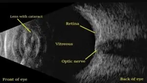
A-Scan biometry is a specialized ultrasound technique used in ophthalmology to measure the axial length (AL) of the eye. It is essential for determining the power of intraocular lenses (IOLs) before cataract surgery and for diagnosing certain eye conditions. This non-invasive procedure provides precise data that helps ophthalmologists plan surgical interventions and assess eye health.
How to work –
A-Scan biometry operates using high-frequency ultrasound waves that travel through the eye and reflect off various structures. The system measures the time taken for these echoes to return, calculating distances between key eye components. The process
involves:
1. Transmitting ultrasound waves through a probe placed on or near the eye.
2. Receiving echoes from different structures like the cornea, anterior chamber, lens, vitreous humor, and retina.
3. Analyzing data to measure the axial length and help determine the appropriate intraocular lens power.
Types of A-Scan-
There are two main types of A-Scan biometry used in clinical practice:
– Contact A-Scan Biometry: The probe touches the cornea directly. Requires topical anesthesia to minimize discomfort.
Quick and widely used but may lead to slight measurement errors due to corneal compression.
– Immersion A-Scan Biometry: The probe is placed in a saline-filled shell without direct contact with the cornea. More accurate because it prevents corneal compression. Preferred for precise intraocular lens calculations.
Clinical Uses of A-Scan Biometry-
A-Scan biometry is crucial in several ophthalmic procedures and conditions, including:
1. Cataract Surgery: Used to determine the correct IOL power, ensuring clear postoperative vision. Helps prevent refractive errors after surgery.
2. Refractive Surgery (LASIK, PRK, etc.): Provides essential preoperative measurements to assess eye structure. Helps determine
eligibility for surgery.
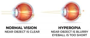
3. Diagnosis of Eye Disorders: Used to detect extreme myopia (nearsightedness) or hyperopia (farsightedness). Helps in identifying ocular conditions like retinal detachment.
4. Glaucoma Monitoring: Can be used to track changes in eye size, which may be associated with certain types of glaucoma.
5. Pediatric Ophthalmology: Helps diagnose congenital eye conditions. Used in cases of microphthalmia (abnormally small eyes) or buphthalmos (enlarged eyes due to high intraocular pressure).
Advantages of A-Scan Biometry-
Non-invasive and painless procedure
Quick and cost-effective compared to advanced imaging techniques. Provides accurate measurements for cataract and refractive surgery planning. Widely available in ophthalmology clinics and hospitals.
Limitations –
Despite its benefits, A-Scan biometry has some limitations: Dependence on Operator Skill: Accuracy depends on proper probe placement.
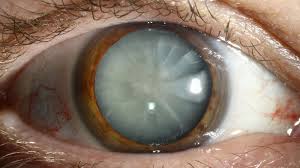
Corneal Compression (in Contact Biometry): Can lead to slight measurement errors. Less Effective in Dense Cataracts: Ultrasound waves may not pass through extremely opaque lenses effectively.
B-Scan
Introduction-
B-Scan ultrasonography is a non-invasive imaging technique used in ophthalmology to visualize the internal structures of the eye and orbit. It is
especially useful when media opacities such as dense cataracts, vitreous hemorrhage, or corneal scars obstruct direct visualization of the retina and posterior segment. B-Scan provides a real-time, cross-sectional view of the eye, assisting in the diagnosis of various ocular and orbital conditions.
How to work –
B-Scan ultrasonography utilizes high-frequency sound waves (8–10 MHz) that penetrate the eye and reflect off different internal structures. The returning echoes are processed by a computer to generate a two-dimensional grayscale image.
Procedure:
1. A water-based gel is applied to the closed eyelid to enhance sound wave transmission.
2. The probe (transducer) is gently placed over the eyelid.
3. The ultrasound waves pass through the eye, and reflected signals create a real-time image on a screen.
4. The ophthalmologist adjusts probe positioning and angles to obtain different cross- sectional views. Unlike A-Scan, which provides a one-dimensional amplitude waveform, B-Scan generates a detailed two-dimensional image of intraocular and orbital structures.
Clinical Uses of B-Scan Ultrasonography –
1. Retinal and Vitreous Disorders-
B-Scan is crucial for diagnosing conditions affecting the posterior segment when direct visualization is obstructed.
– Retinal Detachment (RD): Helps differentiate between total and partial detachment and identifies associated hemorrhages.
– Vitreous Hemorrhage: Detects bleeding within the vitreous cavity, common in diabetic retinopathy, trauma, or retinal tears.
– Vitreous Opacities & Floaters: Identifies inflammatory debris, infections, or posterior vitreous detachment (PVD).
2. Ocular Trauma and Foreign Body Detection B-Scan is highly effective in assessing eye injuries and detecting: Intraocular foreign bodies (IOFB) that may not be visible in routine examinations. Globe integrity in cases of suspected globe rupture or penetrating trauma. Choroidal detachments or hemorrhages caused by blunt trauma.
3. Ocular and Orbital Tumors
– Choroidal Melanoma: Helps determine tumor size, shape, and location.
-Retinoblastoma: Essential for diagnosing childhood intraocular cancer.
– Metastatic Ocular Tumors: Detects tumors that spread from other body organs (e.g., breast or lung cancer).
– Orbital Tumors: Assesses tumors in the eye socket, such as optic nerve gliomas or meningiomas.
4. Evaluation of the Optic Nerve and Glaucoma
– Optic Nerve Head Cupping: Helps assess damage due to advanced glaucoma.
– Optic Nerve Drusen: Identifies deposits in the optic nerve head that can mimic swelling.
5. Diagnosis of Inflammatory and Infectious Conditions
– Endophthalmitis: Detects intraocular inflammation or pus collections in severe infections.
– Posterior Scleritis: Reveals characteristic “T-sign”, indicating fluid accumulation in the posterior sclera.
– Uveitis and Choroiditis: Identifies inflammatory changes in the choroid and retina.
6. Thyroid Eye Disease (Graves’ Orbitopathy) B-Scan helps evaluate extraocular muscle thickening and orbital tissue inflammation, key indicators of thyroid-related eye disease.
7. Pediatric Ophthalmology Applications
– Congenital Cataracts: Helps assess the posterior segment in infants with cloudy lenses.
– Microphthalmia and Anophthalmia: Measures globe size in underdeveloped or absent eyes.
Advantages of B-Scan Ultrasonography-
Non-invasive and painless procedure. Quick and efficient imaging technique.
Safe for all age groups, including pregnant women and children. No exposure to ionizing radiation. Effective in detecting intraocular tumors, retinal detachments, and foreign bodies. Can be performed in emergency settings for trauma evaluation.


