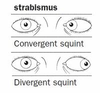
Divergence paralysis or simple stated as paralysis of divergence is demonstrated as the condition by which the distant objects appear to be double. The aspect of diplopia, which is entirely present with the eyes in a medical horizontal plane does not increase when the test object is properly moved within either side. The researchers also stated that the double vision is homonymous as well as it is also less evident amid the approachable near point.
The aspect of divergence paralysis or divergence insufficiency is stated as esotropia or high esophoria at a distance with much lower esophoria or near to normal fixation. It is also stated that primary divergence insufficiency does not possess any other neurological symptoms as well as signs. On the contrary, secondary divergence paralysis possesses the signs as well as symptoms regarding neurologic dysfunction. The neurologic dysfunctions, which would cause diplopia consist of sixth cranial nerve paresis as well as paralysis due to head trauma along with diabetes. The aspect of neurologic dysfunctions is also caused due to the increment of intracranial pressure as well as vascular disease. The patients with the divergence paralysis that possess a pattern of esotropia are properly treated surgically and if they do not respond to or does not desire the prism therapy. The surgical treatment regarding divergence paralysis esotropia is also approached by different techniques, which leads to providing a satisfactory result.
The aspect of diplopia is generated with the paralysis of divergence and it is occurred due to the lack of divergence within the eyes that normally occurs when the entire object is viewed at a distance. This kind of lack of divergence does not occur due to the weakness of the external rectus muscles. It is also stated that rectus muscles do not provide any apparent impairment of the proper function concerning the Divergence Paralysis.
The context of divergence paralysis is entirely characterised by the acquired horizontal homonymous diplopia that leads to the view of distant objects without any limitations of the ocular movement. Divergence paralysis also leads to raised intracranial pressure and it is demonstrated as the aetiological factor within the production of divergence paralysis. There will be much diversity of opinion to describe the underlying mechanism. Paralysis of divergence also presents with varying degrees of severity that is ranging from entire paralysis to the subtle forms of weakness. A wide spectrum of symptoms is noticeable to provide a classic presentation that includes sudden onset of concomitant esotropia with the aspect of diplopia. Moreover, the patients also complain of blurred vision rather than the context of diplopia. The patients with the divergence paralysis lead to illustrate different typical features that will be represented in an appropriate manner. Divergence paralysis is also stated to be an absence of the brainstem signs also provide a rare clinical sign, which is characterised by the esotropia with the distance fixation. This is able to describe with the help of normal abduction that provides no ocular misalignment at the near fixation.
The acute onset of diplopia is demonstrated regarding the increment of distances, which is also stated to be the only complaint. Diplopia that is occurred due to the divergence paralysis is also described in different neurologic diseases but it does not reflect upon meningitis. The aspect of divergence paralysis is entirely described in patients with encephalitis, use of diazepam, syphilis, Fisher syndrome, brainstem hematoma, neuroborreliosis, brainstem ischemia, raised intracranial pressure and many more. The entire duration of divergence palsy within the reversible causes ranges from hours after withdrawing diazepam to days after the treatment that is raised due to intracranial pressure. A particular anatomic site for the divergence centre also remains speculative as well as the most documented clinical cases are entirely relatable to the aspect of brainstem lesions. The support that is provided by neuroradiologic regarding the supportable anatomic localisation for the divergence centre in the brainstem is also acquired within a patient with the small hematoma in tegmentum in relation to upper pons as well as the upper medulla.


