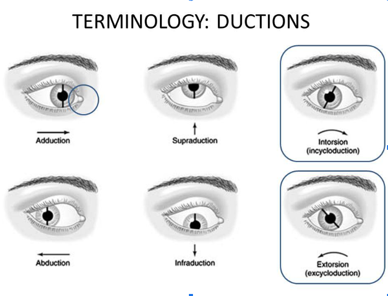
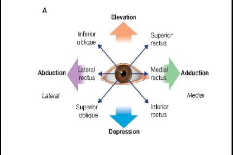
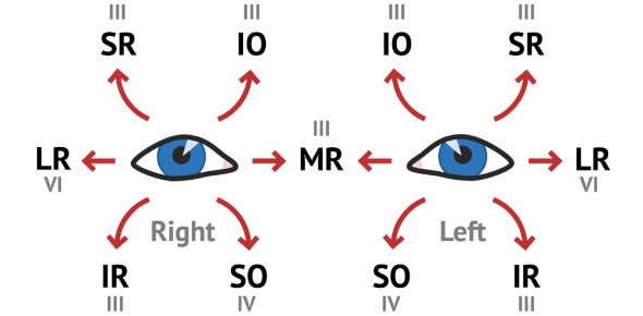
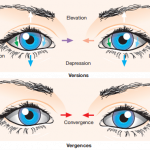
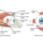
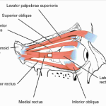
A duction is an eye movement involving only one eye.There are generally six possible movements depending upon the eye’s axis of rotation:
- Adduction: An inward movement ( medial rotation) along the vertical axis.
- Abduction: An outward movement ( lateral rotation) along the vertical axis.
- Supraduction (sursumduction): An upward movement (elevation) along the horizontal axis.
- Infraduction (deosursumduction) : A downward movement ( depression) along the horizontal axis.
- Incycloduction (intorsion) : A rotatory movement along the anteroposterior axis in which superior pole of the cornea (12 o’clock point) moves medially.
- Excycloduction (extortion): A rotatory movement along the anteroposterior axis in which superior pole of the cornea ( 12 o’clock point) moves laterally.
DUCTION TEST
Duction (monocular) testing helps to differentiate between an incomitant deviation due to paresis/paralysis and one due to mechanical restriction. With an incomitant deviation due to a paresis, an underaction seen during version (binocular) testing is less obvious during duction testing, and any overaction will not be seen monocularly. An underaction that is similar when tested monocularly and binocularly suggests a mechanical restriction and may require further testing using forced ductions. To aid or confirm a diagnosis of an underaction/overaction, to measure the extent of the deviation and to assess the degree of incomitancy, further tests are required such as the 9-point cover test.
Forced Duction Test
The forced duction test is performed in order to determine whether the absence of movement of the eye is due to a neurological disorder or a mechanical restriction.
The anesthetized conjunctiva is grasped with forceps and an attempt is made to move the eyeball in the direction where the movement is restricted. If a mechanical restriction is present, it will not be possible to induce a passive movement of the eyeball.
ASSESSMENT OF DUCTION
1.DUCTION TEST: Ductions are monocular movements and are measured at near distance. When examining Ductions, one eye is covered and the fellow eye fixates a spotlight which is moved to bring the fixating eye to the farthest possible position, in all the cardinal directions of gaze. For interpretation of the observations, following methods are in vogue:
- In most frequent practice,the examiner observes whether movement lags or is excessive in any direction. If no lags are noticed, the Ductions are recorded as full, if lags are noticed, the muscle and the eye involved are indicated. Usually,a subjective assessment is made on scale of 7 points (+3 to -3) or 9 points (+4 to -4) . Further , a note is also made of the occurrence of any nystagmoid movements in the presence of full Ductions.
- Judging the normalcy of adduction and abduction in relation to fixed points. Following useful guidelines have been suggested:
- In maximal adduction: An imaginary vertical line through the lower lacrimal punctum should coincide with a boundary line between the inner one-third and the outer two-third of the cornea.
- In excessive adduction, more cornea is hidden.
- In defective adduction, more cornea is visible. Some of the sclera may also be visible.
- In maximal abduction: The lateral limbus touches the outer canthus.
-
- In excessive abduction, some of the cornea is hidden under the outer canthus.
- In defective abduction, some of the sclera is visible between the outer canthus and the limbus.
2.Kestenbaum’s limbus test of motility: The duction movements are measured with the help of a transparent ruler as follows:
- Adduction is measured by noting a difference between the position of the temporal limbus in primary position and maximum adduction
- Abduction is measured by noting a difference between the position of the nasal limbus in primary position and maximum abduction.
- Similarly elevation and depression are measured with respect to inferior limbus and superior limbus, respectively.
- Normal values reported are:
- Adduction: 10 mm
- Abduction: 10mm
- Elevation: 5-7mm
- Depression: 10 mm
3.Subjective perimeter method of measuring Ductions: In this method, to measure the amplitude of duction movements, the patients head is placed into chin rest of a perimeter in such a way that the eye to be examined is in the centre of the perimeter arc or perimeter hemisphere. The other eye is occluded and the patient is asked to fixate and follow the perimeter target that is moved from the centre of the field to periphery. He/she is instructed to indicate, when he/ she can no longer see the target. This point indicates the limit of the Duction movement in that particular direction. Normal values reported by this method are:
– Adduction: 50°
– Abduction: 50°
– Elevation: 40°
– Depression: 50°
- Objective perimeter method or corneal reflex method of measuring Ductions: The amplitude of duction movements can be checked somewhat more objectively by using corneal light reflex. In this method, after closing one eye, patient is asked to turn his/ her eye maximally in a given direction. Then the examiner moves a small flash light along the arc of the perimeter until the reflex from the patients cornea appears to be centred in the pupil. The examiner views it with one eye from the position of flash light. This point gives the limit of particular Duction movement.

