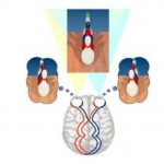
Aniseikonia is an ocular condition, where the image projected to the visual cortex from two retinaie are abnormally unequal in size and shape, mainly aniseikonia is a defect of binocular vision,
The term aniseikonia originates from greek word aniso (unequal) and ekon (image). Mainly 20-30% of people who wearing spectacles, they may have a measurable amount of aniseikonia. The greek word aniseikonia means “Unequal image” and aniseikonia was discussed in 1875, mainly it arises from physiological, retinal and optical cause. Around 20-30% of the general spectacle wearing population may have a measurable amount of aniseikonia within 20-30persent only 5-6% are clinically significant. There is no prevalence for gender, Unequal images can be caused by various cause, mainly
- Optical magnification
- Retinal receptor distribution
Mainly optical magnification mean different retinal image sizes in both eye, Where retinal receptor distribution is different sampling of the retinal images,
Aniseikonia is associated anisometropia, and optical correction of aniseikonia,
Types:
Mainly two types of aniseikonia are seen in clinical practice —
- Static aniseikonia
- Dynamic aniseikonia. or optical induced aniseikonia.
Most commonly aniseikonia occur eye surgery(cataract), When a patient with hypermature cataract in one eye, and he/she did cataract surgery to minimize the refractive error in the operated eye, but other eye require high refractive correction to cure vision, another if a patient have done his/her cataract surgery but implant IOL power is wrong, then also aniseikonia produced.
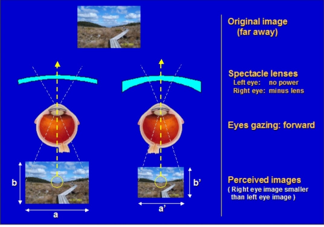
- Static Aniseikonia: Static aniseikonia is the most common type of aniseikonia, mainly it causes eyeglasses, it also associated anisometropia, sometimes aniseikonia will provide double, blur vision, so we cannot determine the real image, where they will provide blur vision, Its also look like 3D movie without wearing 3D glass. In static aniseikonia where our eye moves in same direction then the size of the image is different in each eye.
Example: If a patient with static aniseikonia then, the right image is persived approximately 5% larger then the left eye image, So the aniseikonia is corrected by minify the image in the left eye, approximately 5%, mainly one lens makes left eye vision crystal clear and another lens make right eye vision crystal clear, In this position the patient unable to short it out. Mainly when we solve the anisometropia, then a monocular approch induced aniseikonia and its make the image different in each eye. Normally spectacle induced aniseikonia produced a differences in ocular images in both, and also imposes constant changes in the oculomotor relationship between the two eyes during conjunctive excursions.
2. Dynamic aniseikonia or optical induced anisophoria: Dynamic aniseikonia means that the eye have to rotate different amount of gaze, the aim to look the sharpest vision, Mainly vertical direction eye movement is very important. The cause of unequal image in size may be optical, Its due to different refractive error in two eyes. Dynamic aniseikonia may be congenital, when unequal disposition of the rod and cones in the retina, Optical aniseikonia is a types of aniseikonia due to physical measure difference in size of the retinal image. It arises from uncorrected axial anisometropia. Maddox rod is used to measure the dynamic aniseikonia, Approximately a small vertical phoria is present in primary gaze in all condition.
In dynamic aniseikonia when glasses force the eye to move up- and down or laterally in different speed, So that visual displacement occurs between right eye and left eye. So both image are overlap due to variable size of the image of two eyes. Spectacle and contact lenses are use to correct the aniseikonia.

