Imagine waking up one day to find that the world feels slightly off. The ground seems tilted,
words on a page appear to double or move, and you feel disoriented in the grocery store
because you can't judge distances anymore. For millions of people who experience a traumatic
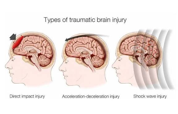
brain injury (TBI) each year, this is not imagination, it is a new reality. While we often think of
brain injuries affecting memory, movement, or speech, the visual system is frequently the
hardest hit. In fact, according to the Neuro-Optometric Rehabilitation Association (NORA), a
staggering 90% of traumatic brain injuries result in vision problems . This is where a specialized
field known as neuro-optometric rehabilitation steps in to rebuild the bridge between the eyes
and the brain.
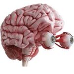
Vision is the brain's ability to interpret what our eyes are seeing. After a TBI, the eyeballs
themselves are often perfectly healthy. The problem however, lies in the brain stem and the
processing centers that integrate the visual information coming in from both eyes . The delicate
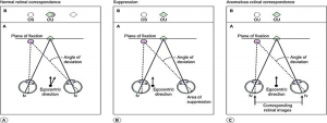
balance of the twelve muscles controlling eye alignment gets disrupted, leading to a "loss of
synchronization". This results in a host of confusing symptoms that standard prescription
glasses simply cannot fix.
The symptoms of TBI-related visual dysfunction are surprisingly diverse. A patient might
experience intermittent or constant double vision, especially when trying to read or sustain
focus . Others suffer from debilitating headaches, fatigue, and a loss of concentration during
visual tasks. Depth perception can vanish, many experience a loss of peripheral vision or a
sensation that the environment is shifting or moving when it is not. Perhaps one of the most
overlooked symptoms is the effect on balance and posture. When the visual system sends
faulty spatial information to the brain, this is a condition known as "visual midline shift
syndrome" (VMSS).
This is where the neuro-optometric rehabilitation optometrist comes in. These specialists are
trained to look beyond the standard eye chart. Unlike a traditional eye exam that might take 30
minutes, a neuro-optometric evaluation is an exhaustive deep dive, often taking two to four
hours over multiple visits. The doctor performs a "21-point probe battery" to assess the
oculomotor system, how the eyes tracks, jump between targets, and work together as a team.
They assess how the brain processes information and how visual dysfunction might be affecting
the patient's gait and posture. Because brain injuries are complex, these optometrists often
work within an interdisciplinary team, coordinating with neurologists, physical therapists, and
occupational therapists to treat the body as a whole.
Once diagnosed, the treatment plan is highly individualized. Contrary to what some might
think, it rarely involves "building up" the eye muscles, because the muscles are not the issue,
the timing and coordination signals from the brain are. Instead, the therapy relies on the brain's
incredible ability to rewire itself, known as neuroplasticity . One of the primary tools in the
toolkit is the use of specialized lenses. While standard glasses correct for clarity, neuro-
optometry uses therapeutic prisms and yoked prisms (prisms that shift the entire field of vision
in a specific direction) to help realign the visual world, reduce double vision, and even improve
balance and spatial awareness .
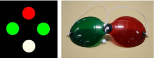
In addition to lenses, in-office vision therapy involves a series of structured activities designed
to retrain the brain. This might include working with "vectograms," polarized images that train
the eyes to converge and diverge efficiently . For patients struggling with focusing, therapists
use plus and minus lenses to "exercise" the eye's focusing system . To rebuild peripheral
awareness, a patient might be asked to focus on a central chart while simultaneously
identifying colors appearing in their side vision . For those with oculomotor dysfunction, a
rotating pegboard can help retrain smooth eye movements and hand-eye coordination. The
goal of these exercises is to create new neural pathways.
While the field is growing, it is not without controversy. Some medical professionals,
particularly neuro-ophthalmologists, note that the scientific evidence base for specific
diagnoses like "post-trauma vision syndrome" relies heavily on case reports rather than large-
scale clinical trials . However, there is a growing consensus that for oculomotor impairments
following TBI, restorative approaches are beneficial . Many occupational therapists are now
integrating vision therapy techniques into their practices, recognizing that you cannot address a
patient's mobility or daily living skills without first addressing how they see and interact with
their environment.
For anyone living with the aftermath of a brain injury, it is vital to know that visual problems are
often overlooked in initial treatment, with symptoms sometimes appearing months after the
event. If you or a loved one experiences headaches, dizziness, balance issues, or visual
confusion following a head injury, it is worth seeking out a specialist. Early diagnosis is key, and
organizations like NORA (Neuro-Optometric Rehabilitation Association) can help locate a
certified provider . The journey of neuro-optometric rehabilitation is a powerful reminder that
the brain is not a static organ; it is a dynamic, plastic system capable of remarkable healing
when guided by the right therapy.