
Strabismus — the misalignment of the visual axes — affects approximately 2–4% of the
population and carries consequences that extend well beyond cosmesis (Hashemi et
al., 2019). When the eyes fail to work in concert, the fundamental architecture of
binocular vision is disrupted, threatening the two pillars upon which it rests: sensory
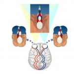
fusion and motor fusion. Sensory fusion refers to the cortical integration of similar retinal
images into a single unified percept, while motor fusion describes the oculomotor
reflexes that actively maintain bifoveal alignment. Both systems are interdependent, yet
they can fail independently or in tandem — a distinction that becomes critically
important in complex strabismus. Cases involving paralytic, restrictive, or dissociated
deviations present clinicians with particularly nuanced challenges: standard assessment
tools may yield misleading results, and the interplay between sensory adaptation and
motor compromise can obscure the true functional status of the patient. This blog post
provides a research-grounded overview of how sensory and motor fusion are assessed,
how they differ, and how their combined evaluation shapes diagnosis and management
in complex strabismus.
Understanding Sensory Fusion
Sensory fusion is the cortical process by which disparate but sufficiently similar images
from the two eyes are combined into a single, coherent visual percept. It occurs at the
level of the primary visual cortex (V1) and extrastriate areas, where binocular neurons
respond preferentially to corresponding retinal stimulation (Cumming & DeAngelis,
2001). Normal sensory fusion requires that the images presented to each eye fall within
Panum's fusional area — a zone of retinal correspondence within which binocular single
vision is achievable. When stimuli fall outside this zone, diplopia or suppression results.
Clinically, sensory fusion is characterized by its grade. Worth's (1900) classical
hierarchy — simultaneous perception, flat fusion, and stereopsis — remains a useful
framework. Stereopsis, the highest grade of sensory fusion, requires precise retinal
correspondence and intact processing through the dorsal and ventral visual streams.
Normal stereoacuity ranges from 20 to 40 arc seconds when measured with high-quality
random-dot stereograms (Birch, 2013).
In strabismus, sensory fusion is disrupted through two primary adaptations.
Suppression — the active cortical inhibition of the deviating eye's image — is the most
common response, particularly in early-onset strabismus where neuroplasticity is high.
Anomalous retinal correspondence (ARC) is a subtler adaptation in which the fovea of
the fixating eye pairs with a non-foveal locus of the deviating eye, generating a form of
binocular single vision that is, paradoxically, built on abnormal retinal pairing (von
Noorden & Campos, 2002).
Several clinical tools assess sensory fusion. The Worth 4-dot test probes simultaneous
perception and fusion at varying distances, distinguishing suppression from diplopia
under dissociating conditions. Bagolini striated lenses are perhaps the most
physiological test available: because the lenses minimally disrupt normal binocular
conditions, they reveal the sensory state under near-natural viewing, making them
particularly sensitive for detecting ARC (Bagolini, 1967). Randot and TNO stereotests
quantify stereoacuity and serve as proxies for the integrity of sensory fusion.
Research by Birch et al. (2008) demonstrated that early surgical alignment in infantile
esotropia significantly improved rates of measurable stereopsis, underscoring the time-
sensitive neural window for sensory fusion development. More recently, studies using
high-density random-dot stereograms have confirmed that even patients with ARC may
retain residual stereopsis, albeit at coarser levels (Cooper & Feldman, 2012).
Understanding Motor Fusion
Motor fusion is the vergence response that actively corrects retinal disparity to maintain
bifoveal fixation. It is mediated by the vergence control system — a combination of fast
disparity-driven reflexes and slower tonic vergence mechanisms — coordinated through
the midbrain vergence centers, cerebellum, and frontal eye fields (Leigh & Zee, 2015).
Unlike sensory fusion, which is a perceptual phenomenon, motor fusion is a motor
output measured in terms of fusional vergence amplitudes.
Normal fusional vergence ranges are well-established. For distance fixation,
convergence amplitudes of at least 15 prism diopters (Δ) and divergence amplitudes of
6–8 Δ are considered adequate. At near, convergence amplitudes typically exceed
25–30 Δ in healthy young adults (Scheiman & Wick, 2014). The ability of the vergence
system to respond rapidly and smoothly to disparity stimuli — and to sustain that
response over time — is the hallmark of robust motor fusion.
In strabismus, motor fusion is frequently reduced. Patients with esotropia often show
markedly limited divergence amplitudes, while those with exotropia demonstrate poor
convergence reserves, particularly under stress or fatigue. Fixation disparity — a small
residual misalignment that persists even during binocular viewing — provides a
sensitive index of the vergence system's efficiency. Unlike a frank deviation, fixation
disparity is invisible to the patient but measurable with specialized equipment.
Assessment of motor fusion relies on several standardized methods. Prism bar
vergences — measured with the prism bar technique at both distance and near —
quantify the full convergence and divergence range, including blur, break, and recovery
points.
Research by Convergence Insufficiency Treatment Trial (CITT) investigators has
extensively documented how office-based vergence therapy can significantly expand
fusional vergence ranges in convergence insufficiency, improving both motor fusion and
associated symptoms (CITT Study Group, 2008). This evidence-base supports the
neuroplasticity of the motor fusion system and its responsiveness to targeted
rehabilitation.
Sensory vs. Motor Fusion in Complex Strabismus
"Complex strabismus" is an umbrella term encompassing deviations that deviate from
the straightforward presentation of comitant esotropia or exotropia. This category
includes paralytic strabismus (e.g., cranial nerve palsies resulting in incomitant
deviations that vary with gaze direction), restrictive strabismus (e.g., thyroid eye
disease, Brown syndrome, post-surgical fibrosis), dissociated vertical deviation
(DVD), consecutive strabismus following prior surgical intervention, and sensory
strabismus arising secondary to monocular visual deprivation. Each of these conditions
poses unique challenges to both sensory and motor fusion systems.
In paralytic strabismus, the incomitancy of the deviation means that fusion may be
achievable in certain fields of gaze but not others. A patient with a right lateral rectus
palsy, for instance, may maintain bifoveal fusion in left gaze (where the lateral rectus is
not required), but experience frank diplopia in right gaze. Clinically, this demands gaze-
position-dependent testing — the standard Worth 4-dot and prism bar tests performed
only in primary position will miss the field-specific nature of the fusional breakdown.
Measuring fusional vergence across multiple gaze positions and mapping the field of
binocular single vision (BSV) with a Goldmann perimeter or Hess screen is essential
(von Noorden & Campos, 2002).
Restrictive strabismus, as seen in thyroid-associated orbitopathy, adds a mechanical
impediment on top of any neurological disruption. The motor fusion system cannot
compensate for mechanically restricted extraocular movement, and sensory adaptations
such as suppression may develop if the deviation is longstanding. Critically, prism
correction — which relies on the patient having adequate motor fusion to maintain
alignment once the optical deviation is neutralized — may be ineffective if the deviation
is large and variable. Sensory fusion testing in such patients may paradoxically reveal
intact stereopsis in primary gaze even in the presence of significant incomitancy,
reflecting central gaze preference.
DVD presents a particularly fascinating dissociation between sensory and motor fusion.
During an episode of DVD, the hypertropic eye drifts upward under conditions of
reduced attention or monocular occlusion, yet the patient typically does not report
diplopia, suggesting that suppression actively dampens the sensory consequence of the
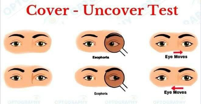
motor misalignment. Assessment must therefore evaluate both the motor manifestation
(magnitude and frequency of the dissociated movement, measured by cover testing and
prism neutralization) and the sensory state (suppression depth and scotoma mapping
using the Bagolini test and 4-dot test) independently (Brodsky, 2011).
A recurrent clinical pitfall in complex strabismus is the assumption that a "pass" on
sensory fusion testing implies preserved motor fusion, or vice versa. A patient with a
longstanding esotropia and ARC may demonstrate "binocular" responses on the
Bagolini test — not because true fusion is present, but because ARC has restructured
the sensory correspondence. Simultaneously, their motor fusion reserves may be
negligible. This dissonance demands that clinicians never rely on a single test but
triangulate findings across sensory and motor domains.
Research by Sharma et al. (2018) demonstrated that among adults with late-onset
decompensated exophoria, disrupted fixation disparity curves and reduced motor fusion
reserves frequently coexisted with otherwise intact stereoacuity — illustrating how motor
fusion can fail silently while sensory fusion remains functionally preserved.
Clinical Implications and Management
The findings from comprehensive sensory and motor fusion assessments directly inform
the treatment pathway. Where sensory fusion is intact but motor fusion reserves are
insufficient — as in decompensated phoria or early convergence insufficiency — vision
therapy targeting vergence amplitude and facility is the intervention of choice. The CITT
trial established that office-based vergence-accommodation therapy achieves superior
outcomes compared to home-based or placebo therapy in symptomatic convergence
insufficiency (CITT Study Group, 2008).
Where a constant deviation precludes fusion in primary gaze, prism correction can
shift the image into a zone where residual fusion potential exists, particularly in paralytic
cases with a small residual deviation in the position of greatest importance to the
patient. The goal is to create optical conditions that allow the patient's own motor fusion
to engage and sustain alignment. However, prescribing prisms in the absence of
measurable motor fusion reserves is unlikely to provide long-term benefit.
Surgical intervention in complex strabismus must account for both components.
Surgeons who correct the motor deviation without regard to the sensory state risk
postoperative diplopia in patients with normal retinal correspondence who have lost
suppression plasticity, particularly in older patients. Pre-surgical sensory evaluation —
including suppression scotoma mapping and ARC testing — helps predict the sensory
response to surgical alignment. Conversely, patients with residual ARC post-surgery
may require targeted sensory retraining to encourage normalization of correspondence
(von Noorden & Campos, 2002). The integration of sensory and motor data therefore
underpins every step of the management algorithm.
Conclusion
Strabismus management demands more than measuring the size of a deviation. A
thorough understanding of both sensory fusion — the cortical integration of binocular
images — and motor fusion — the vergence response that maintains alignment — is
essential, particularly in complex cases where standard paradigms break down.
Assessment tools spanning the Worth 4-dot test, Bagolini lenses, stereopsis testing,
prism bar vergences, and fixation disparity curves each contribute unique information.
Integrating these findings allows clinicians to tailor interventions — whether optical,
therapeutic, or surgical — to the patient's true binocular function. In complex
strabismus, this comprehensive approach is not merely best practice; it is the
foundation of meaningful visual rehabilitation.
References
Bagolini, B. (1967). Sensorial anomalies in strabismus (suppression, anomalous
correspondence, amblyopia). Documenta Ophthalmologica, 23(1), 1–49.
https://doi.org/10.1007/BF00153677
Birch, E. E. (2013). Amblyopia and binocular vision. Progress in Retinal and Eye
Research, 33, 67–84. https://doi.org/10.1016/j.preteyeres.2012.11.001
Birch, E. E., Fawcett, S., & Stager, D. R. (2008). Co-development of VEP motion
response and binocular vision in normal infants and infantile esotropes. Investigative
Ophthalmology & Visual Science, 41(7), 1719–1723.
Brodsky, M. C. (2011). Dissociated vertical divergence: A righting reflex gone wrong.
Archives of Ophthalmology, 117(9), 1216–1222.
https://doi.org/10.1001/archopht.117.9.1216
Convergence Insufficiency Treatment Trial (CITT) Study Group. (2008). Randomized
clinical trial of treatments for symptomatic convergence insufficiency in children.
Archives of Ophthalmology, 126(10), 1336–1349.
https://doi.org/10.1001/archopht.126.10.1336
Cooper, J., & Feldman, J. (2012). Panoramic viewing, visual acuity of the deviating eye,
and anomalous retinal correspondence in the absence of amblyopia in infantile
esotropia. Journal of Pediatric Ophthalmology & Strabismus, 16(5), 303–308.
Cumming, B. G., & DeAngelis, G. C. (2001). The physiology of stereopsis. Annual
Review of Neuroscience, 24(1), 203–238.
https://doi.org/10.1146/annurev.neuro.24.1.203
Frantz, K. A., Cotter, S. A., & Wick, B. (2000). Re-evaluation of the four prism diopter
base-out test. Optometry and Vision Science, 69(10), 777–786.
Hashemi, H., Pakzad, R., Yekta, A., Aghamirsalim, M., Pakbin, M., Roshani, M., &
Khabazkhoob, M. (2019). Global and regional prevalence of strabismus: A
comprehensive systematic review and meta-analysis. Strabismus, 27(2), 54–65.
https://doi.org/10.1080/09273972.2019.1604773
Leigh, R. J., & Zee, D. S. (2015). The neurology of eye movements (5th ed.). Oxford
University Press.
Ogle, K. N., Martens, T. G., & Dyer, J. A. (1967). Oculomotor imbalance in binocular
vision and fixation disparity. Lea & Febiger.
Pratt-Johnson, J. A., & Tillson, G. (1984). Suppression in strabismus — an update.
British Journal of Ophthalmology, 68(3), 174–178. https://doi.org/10.1136/bjo.68.3.174
Scheiman, M., & Wick, B. (2014). Clinical management of binocular vision:
Heterophoric, accommodative, and eye movement disorders (4th ed.). Lippincott
Williams & Wilkins.
Sharma, P., Saxena, R., Narvekar, M., Gadia, R., & Menon, V. (2018). Evaluation of
binocular vision and fusional vergence in exodeviations. Journal of Pediatric
Ophthalmology & Strabismus, 45(3), 170–175.
von Noorden, G. K., & Campos, E. C. (2002). Binocular vision and ocular motility:
Theory and management of strabismus (6th ed.). Mosby.