The lens of the eye plays a critical role in focusing light on the retina, facilitating clear vision. When thelens is improperly positioned due to a partial dislocation, the condition is referred to as “subluxation of the lens.” This condition is medically significant as it can lead to vision impairments and other complications. Understanding the causes, symptoms, diagnosis, and management options of lens subluxation is crucial for both eye care professionals and patients.
1. Lens Subluxation:-
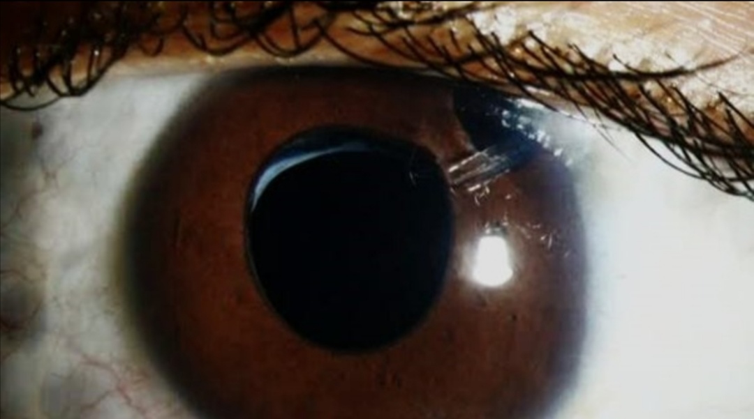
Subluxation of the lens, also known as “ectopia lentis,” occurs when the lens is partially displaced from its normal anatomical position within the eye. The condition typically arises from damage or weakening of the zonules—fibers that hold the lens in place. While complete dislocation (luxation) involves the total displacement of the lens, subluxation is characterized by partial dislocation, meaning the lens remains somewhat attached but is no longer properly aligned.
2. Causes of Lens Subluxation-
Lens subluxation can result from various factors, with the most common causes being trauma, genetic conditions, and certain diseases:
a) Trauma
Blunt or penetrating injuries to the eye are among the leading causes of lens subluxation. A strong impact can weaken or sever the zonules, causing
the lens to shift from its natural position. In some cases, the lens may remain subluxated immediately after the trauma, while in other instances, the displacement may occur gradually.
b) Genetic Disorders
Certain inherited disorders are strongly associated with lens subluxation. Among these, *Marfan syndrome* is perhaps the most well-known. This
genetic condition affects connective tissue, leading to a range of systemic abnormalities, including weak zonules in the eye. People with Marfan syndrome have an increased risk of lens subluxation, often in both eyes.
Other genetic conditions that may lead to lens subluxation include:
– Homocystinuria : A metabolic disorder characterized by high levels of homocysteine, which
can cause the zonules to weaken and result in lens displacement.
– Ehlers-Danlos syndrome : A group of disorders affecting connective tissue that may lead to subluxation due to the fragility of zonules.
– Weill-Marchesani syndrome : A rare connective tissue disorder that also impacts the eye, leading to lens subluxation among other ocular complications.
c) Other Conditions
Certain eye diseases, such as pseudoexfoliation syndrome, can lead to the gradual weakening of the zonules. Additionally, advanced cataracts or long- standing glaucoma may also contribute to the loosening of these fibers, resulting in subluxation over time.
3. Symptoms of Lens Subluxation:-
The symptoms of lens subluxation can vary in intensity depending on the degree of displacement and the underlying cause. Some common symptoms
include:
– Blurry Vision : Due to the misalignment of the lens, the eye may struggle to properly focus light, leading to blurry or distorted vision.
– Double Vision : In some cases, patients may experience diplopia, or double vision, especially if the displacement is significant.
– Glare and Halos : Patients may notice increased glare or halos around lights, particularly at night.
– Irregular Astigmatism : The irregular positioning of the lens may cause astigmatism that is difficult to correct with glasses or contact lenses.
– Eye Discomfort : Some individuals with lens subluxation may experience mild discomfort or a sensation of something being “off” in their vision.
4. Diagnosis of Lens Subluxation:-
Accurate diagnosis of lens subluxation requires a thorough eye examination by an ophthalmologist or optometrist. The following diagnostic methods are typically employed:
a) Slit-Lamp Examination
A slit-lamp microscope is a primary tool for examining the front part of the eye, including the lens. Through this examination, the eye care professional can assess the position of the lens, the condition of the zonules, and any associated signs of damage.
b) Ophthalmoscopy
Ophthalmoscopy allows the doctor to examine the back of the eye, including the retina and optic nerve. This can help rule out other ocular conditions that may be contributing to the patient’s symptoms.
c) Ultrasound Biomicroscopy (UBM)
For more detailed imaging of the anterior segment of the eye, ultrasound biomicroscopy may be used. This high-frequency ultrasound provides detailed images of the lens and zonules, allowing the doctor to precisely assess the extent of subluxation.
d) Genetic Testing
If a genetic condition like Marfan syndrome or homocystinuria is suspected, genetic testing may be recommended to confirm the diagnosis. This can
help guide further management and alert the patient to other possible systemic complications.
5. Management and Treatment of Lens Subluxation
The treatment of lens subluxation depends on the severity of the condition, the degree of visual impairment, and the underlying cause. Management options range from non-surgical approaches to surgical interventions.
a) Non-Surgical Management
In cases of mild lens subluxation where vision is only minimally affected, the condition can often be managed conservatively:
– Corrective Lenses : Glasses or contact lenses may be prescribed to improve vision. If astigmatism is present, toric lenses may be required.
– Monitoring : In certain cases, regular monitoring of the eye’s condition is recommended to ensure the subluxation does not worsen or lead to additional complications like glaucoma or cataracts.
b) Surgical Treatment
Surgical intervention is typically required if the subluxation is severe or if non-surgical methods fail to restore adequate vision. Surgical options include:
– Lens Removal and Intraocular Lens (IOL)
Implantation : This is the most common surgery for
severe subluxation. The dislocated lens is removed, and an artificial intraocular lens (IOL) is implanted in its place. In cases where the zonules are severely damaged, special techniques or devices may be used to secure the IOL in place.
– Scleral Fixation of the IOL : In cases of significant zonular weakness, a scleral-fixated IOL may be required. This involves suturing the IOL to the sclera (the white part of the eye) to ensure stability.
– Pars Plana Vitrectomy: This procedure is sometimes performed in conjunction with lens removal, especially if vitreous gel (the gel-like
substance in the center of the eye) is prolapsed or if retinal complications are present.
c) Management of Underlying Conditions For patients with genetic disorders such as Marfan syndrome or homocystinuria, addressing the underlying condition is an essential component of treatment. This may involve systemic medications, lifestyle modifications, or monitoring for other health
complications, such as cardiovascular issues, that are commonly associated with these conditions.
6. Prognosis and Complications:-
The prognosis for patients with lens subluxation varies depending on the underlying cause, the severity of the displacement, and the effectiveness of treatment. When diagnosed early and managed appropriately, many patients can achieve goo visual outcomes.
However, untreated or improperly managed lens subluxation can lead to complications, including:
– Glaucoma : Increased intraocular pressure due to lens displacement can lead to secondary glaucoma.
– Cataract Formation : Prolonged subluxation can contribute to the development of cataracts, further impairing vision.
– Retinal Detachment : In rare cases, severe lens dislocation can increase the risk of retinal detachment, a sight-threatening condition.
Conclusion:-
Subluxation of the lens is a potentially serious ocular condition that requires timely diagnosis and appropriate management. While genetic disorders and trauma are common causes, careful monitoring and treatment can significantly improve the visual and overall health outcomes for affected individuals.


