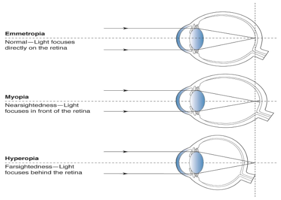
The human eye is remarkably adept at fine-tuning its own optical system. During early
childhood, most infants are born mildly hyperopic (farsighted). Yet, through a finely tuned,
visually driven feedback loop known as emmetropization, the eye precisely scales its axial
length to match the focal power of its optical components. By early childhood, this mechanism
usually delivers crisp, uncorrected distance vision—a homeostatic state called emmetropia.
However, over the past few decades, a dramatic epidemiological shift has disrupted this natural
physiological process. Across the globe—particularly in urbanized centers—eyes are no longer
halting their elongation once clear vision is achieved. Instead, they continue to grow excessively
along their anteroposterior axis, transitioning from healthy emmetropization into myopigenesis,
the chronic cellular and structural pathway toward progressive nearsightedness.
Understanding how the eye shifts from a harmonious visual development trajectory to a
pathological refractive state is one of the most urgent frontiers in modern optometry, ophthalmology, and visual neuroscience.
1. The Precision Mechanism of Emmetropization
Emmetropization is an active, locally regulated biological control system. It relies continuously
on visual input to orchestrate the growth of the sclera, the choroid, and the retina so that
incoming parallel light rays focus precisely onto the photoreceptor layer without dynamic
accommodation.
power over the first years of life, the ocular globe elongates to compensate, maintaining a
delicate balance between focal length and axial dimension.
and optical defocus (blur) and translates these visual inputs into precise biochemical
cascades without requiring direct signal processing from the central brain.
adjusts its thickness to shift the retina toward the focal plane. Simultaneously, the scleral
extracellular matrix remodels its collagen architecture, either slowing down or accelerating
tissue synthesis to modulate axial expansion.
Because this feedback loop operates locally across different retinal regions, individual quadrants
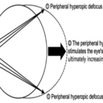
of the eye can direct localized growth. If the eye perceives hyperopic defocus—where the focal
plane falls behind the photoreceptors—it triggers signaling cascades that accelerate scleral
matrix breakdown and axial lengthening. Conversely, myopic defocus—where light focuses in
front of the retina—signals the sclera to inhibit growth, bringing elongation to a controlled halt.
2. The Biological Shift Toward Myopigenesis
Myopigenesis refers to the initiation and cellular progression of structural changes that convert a
homeostatic, emmetropic eye into a myopic one. Rather than stabilizing at a neutral refractive
target, the structural integrity of the posterior segment degrades, allowing sustained posterior
elongation.
What causes this homeostatic feedback system to overshoot its biological stop signal? The
transition from emmetropization to myopigenesis occurs when environmental and optical cues
deceive the retina into perceiving persistent hyperopic defocus, even when central visual acuity
remains sharp.
Key Mechanistic Drivers of Myopigenesis
1. Peripheral Defocus and Lag of Accommodation: During prolonged near tasks, the eye's
crystalline lens frequently under-accommodates relative to the object distance—a
phenomenon termed accommodation lag. This projects hyperopic defocus onto the central
retina. Moreover, conventional single-vision spectacle lenses correct central vision while
casting light far behind the peripheral retina. Recognizing this peripheral hyperopic defocus,
the peripheral retina triggers generalized axial elongation across the entire ocular shell.
2. Contrast and Spatial Frequency Modulation: Modern visual tasks expose the retina to
high-contrast, high-spatial-frequency visual input on digital screens at constant working
distances. Excessive exposure to high-contrast artificial environments alters normal retinal
receptive field dynamics, dampening the naturally occurring biochemical signals that instruct
the sclera to cease growth.
3. Retinal Neurotransmitter Disruption (Dopamine Depletion): Dopamine is a primary
retinal neurotransmitter synthesized by amacrine cells that actively inhibits ocular growth. Its
release is strongly stimulated by high-intensity, full-spectrum outdoor sunlight. Indoor
ambient lighting rarely exceeds 300 to 500 lux, whereas direct outdoor sunlight reaches
10,000 to 100,000 lux. Low indoor illumination leads to chronic retinal dopamine depletion,
effectively removing a vital biological brake on axial expansion.
3. Structural & Cellular Matrix Alterations
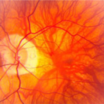
The transition to myopigenesis is not merely an optical nuisance; it involves permanent
structural degradation of the tissues forming the posterior pole of the globe. As axial length
increases, structural biomechanics change significantly across every layer of the posterior
segment.
4. Molecular Signaling Cascades in Ocular Remodeling
At the molecular level, the shift from emmetropization to myopigenesis involves a complex
sequence of chemical messengers traveling from the retina, through the retinal pigment
epithelium (RPE) and choroid, to the sclera:
sclera during hyperopic defocus suppresses collagen synthesis, making the scleral shell
more compliant and susceptible to stretching under intraocular pressure.
fibrils and structural proteoglycans within the scleral matrix, reducing scleral thickness and
dry weight.
as a key intermediate mediator signaling the scleral fibroblasts to reconfigure structural extracellular components.
5. Environmental Acceleration & Genetic Susceptibility
While polygenic inheritance sets baseline susceptibility, environmental exposure dictates
whether myopigenesis takes hold. Genome-wide association studies (GWAS) have identified
over 500 genetic loci linked to refractive error, but genetic drift cannot account for the rapid
global spike in myopia over just two generations.
The Modern Visual Environment Paradox: Children today spend unprecedented hours
indoors engaged in high-demand near vision while deprived of direct sunlight. This
environment combines three powerful myopigenic triggers: high lag of accommodation, low
ambient luminance, and flat spatial surroundings lacking distant optical targets.
small, flat rooms. The absence of long-range focal planes (beyond 6 meters) deprives the
retina of relaxing focal cues, maintaining continuous tonic accommodation.
prolonged reading from books and handheld devices significantly increase the cumulative
duration of hyperopic retinal defocus.
6. Clinical Interventions: Restoring Ocular Homeostasis
Modern clinical myopia management aims to halt the destructive biological pathways of
myopigenesis and recreate the physiological stop signals characteristic of natural
emmetropization.
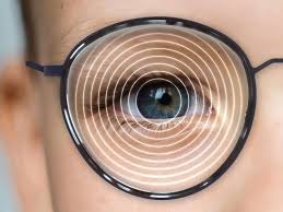
highly specialized spectacle lenses (e.g., DIMS or HAL technology) correct central vision
while introducing simultaneous myopic peripheral defocus. Light focusing in front of the
peripheral retina sends a continuous biological signal to inhibit further axial expansion.
0.05%) bind to muscarinic acetylcholine receptors in the retina and sclera. This inhibits
scleral matrix degradation without impairing daily pupil reaction or accommodation when
properly dosed.
physical activity under natural sunlight boosts endogenous retinal dopamine production,
strengthening the biological barrier against axial growth.
7. A Paradigm Shift in Vision Care and Public Health
The movement from emmetropization to myopigenesis represents a fundamental redefinition of
refractive error in modern medicine. Nearsightedness is no longer viewed as a simple benign
optical inconvenience to be neutralized with standard corrective lenses. Instead, it is recognized
as a complex, environmentally driven physiological dysfunction with significant structural
consequences.
Uncontrolled axial elongation dramatically increases lifetime risks for vision-threatening
pathologies, including myopic maculopathy, retinal detachment, glaucoma, and premature
cataracts. By dissecting the precise cellular signals that push the eye past its natural
emmetropic boundary, researchers and clinicians are transforming vision care from passive
symptom correction into active, preventive disease management—preserving structural ocular
health and visual clarity for generations to come.