Introduction
Dry Eye Disease (DED) is a common, multifactorial
disorder of the ocular surface characterized by a loss
of tear film homeostasis. It presents with symptoms
such as dryness, irritation, burning, foreign body
sensation, and fluctuating vision. According to the
TFOS DEWS II definition, hyperosmolarity,
inflammation, tear film instability, and neurosensory
abnormalities are key contributors.Traditional
diagnostic methods like the Schirmer test, Tear
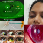
Break-Up Time (TBUT), and ocular surface staining often show variability and poor correlation
with symptoms. In recent years, tear film biomarkers have emerged as objective, sensitive, and
reliable tools for evaluating dry eye and guiding its management.
Tear Film Structure and Function
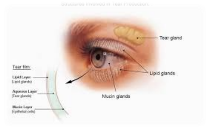
The tear film is a trilaminar structure:
1. Lipid Layer (Outer)
Secreted by meibomian glands
Prevents evaporation
Provides tear film stability
2. Aqueous Layer (Middle)
Produced by lacrimal glands
Contains proteins, enzymes, and electrolytes
Provides nutrition and antimicrobial defense
3. Mucin Layer (Inner)
Secreted by conjunctival goblet cells
Helps tears adhere to the ocular surface
Any disruption in these layers leads to tear film instability and contributes to DED. Biomarkers
help identify which component is primarily affected.
Tear Film Biomarkers in Dry Eye
Biomarkers are measurable substances in tears that indicate physiological or pathological changes.
1. Tear Osmolarity
Considered the gold standard biomarker
Normal: ~275–300 mOsm/L
Dry eye: >308 mOsm/L or inter-eye variability >8 mOsm/L
Significance:
Reflects tear film instability and dehydration
Increased osmolarity damages epithelial cells and triggers inflammation
2. Inflammatory Biomarkers
a. MMP-9 (Matrix Metalloproteinase-9)
Elevated in dry eye
Breaks down epithelial tight junctions
Measured using point-of-care tests (e.g., InflammaDry)
b. Cytokines
Includes IL-1, IL-6, TNF-α
Elevated in moderate to severe dry eye
Indicate immune system activation
3. Lacrimal Gland Function Biomarkers
a. Lactoferrin
Produced by lacrimal glands
Decreased in aqueous-deficient dry eye
Has antimicrobial and anti-inflammatory properties
b. Lysozyme
Protects against bacterial infection
Reduced in dry eye
4. Mucin Biomarkers
MUC5AC is a key mucin protein
Decreased in goblet cell dysfunction
Seen in conditions like vitamin A deficiency and ocular surface disease
5. Lipid Biomarkers
Altered lipid composition in meibomian gland dysfunction (MGD)
Leads to increased evaporation
6. Emerging Biomarkers
Proteomics (tear protein profiling)
Lipidomics and metabolomics
Nerve growth factor (NGF)
These are mainly used in research and advanced diagnostics
Evaluation of Dry Eye Using Tear Film Biomarkers
1. Patient History and Symptom Assessment
Burning, dryness, itching, photophobia
Fluctuating vision
Use of digital devices, contact lenses, medications
Questionnaires
OSDI (Ocular Surface Disease Index)
Helps quantify severity
2. Conventional Clinical Tests
a. Schirmer Test
Measures tear production
<10 mm indicates deficiency
b. TBUT (Tear Break-Up Time)
<10 seconds suggests instability
c. Ocular Surface Staining
Fluorescein, Rose Bengal, Lissamine green
Detect epithelial damage
3. Biomarker-Based Diagnostic Approach
a. Tear Osmolarity Measurement
Devices like TearLab
Quick, non-invasive
High diagnostic accuracy
b. MMP-9 Testing
Detects inflammation
Positive result (>40 ng/mL) indicates active inflammation
c. Lactoferrin Testing
Indicates lacrimal gland function
Low levels suggest aqueous deficiency
d. Tear Protein Analysis
ELISA, mass spectrometry
Detect multiple biomarkers simultaneously
4. Interpretation of Biomarkers
Biomarker Finding Interpretation
High osmolarity >308 mOsm/L Tear instability
High MMP-9 Positive Inflammation
Low lactoferrin Reduced Aqueous deficiency
Low MUC5AC Reduced Goblet cell dysfunction
Lipid changes Abnormal Evaporative dry eye
5. Advantages of Biomarker Evaluation
Objective and reproducible
Detect early disease
Helps classify dry eye subtype
Useful for monitoring therapy
Management of Dry Eye Using Biomarkers
Biomarker-based management allows personalized treatment.
1. General Measures
Environmental modification (avoid dry air, screen breaks)
Proper hydration
Use of humidifiers
2. Tear Substitutes
Artificial tears (preservative-free preferred)
Low osmolarity solutions help dilute inflammatory mediators
Types:
Aqueous-based
Lipid-based (for evaporative dry eye)
Gel and ointments (for severe cases)
3. Anti-inflammatory Therapy
Indicated when inflammatory biomarkers (e.g., MMP-9) are elevated.
Options:
Corticosteroids (short-term use)
Cyclosporine A (chronic inflammation)
Lifitegrast (T-cell inhibition)
4. Osmoprotective Therapy
Reduces hyperosmolar stress
Contains compatible solutes like:
o Trehalose
o Erythritol
o L-carnitine
5. Management of Meibomian Gland Dysfunction
Warm compresses
Lid massage and hygiene
Lipid-based tears
Oral doxycycline (anti-inflammatory effect)
6. Tear Stimulation (Secretagogues)
Increase tear production
Useful in aqueous-deficient dry eye
7. Nutritional Therapy
Omega-3 fatty acids
Improve lipid layer and reduce inflammation
8. Punctal Occlusion
Blocks tear drainage
Used in severe aqueous deficiency
9. Advanced Therapies
Autologous serum eye drops
Platelet-rich plasma (PRP)
Scleral contact lenses
Amniotic membrane therapy
Biomarker-Guided Treatment Strategy
Biomarker Finding Treatment Approach
High osmolarity Artificial tears + osmoprotective agents
Positive MMP-9 Anti-inflammatory therapy
Low lactoferrin Tear substitutes + punctal plugs
Lipid abnormality Lid hygiene + lipid-based tears
Low mucin Lubricants + treat ocular surface disease
Monitoring and Follow-Up
Biomarkers play a crucial role in follow-up:
Tear osmolarity → normalization indicates improvement
MMP-9 → reduction shows decreased inflammation
Lactoferrin levels → improved lacrimal function
Regular monitoring helps:
Adjust treatment
Prevent complications
Improve long-term outcomes