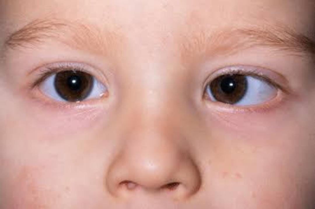
STRABISMUS :Strabismus or squint is a failure of the two eyes to maintain proper alignment. Proper evaluation and timely intervention are necessary to prevent permanent forms of visual dysfunction such as diminished depth perception and lazy eye.
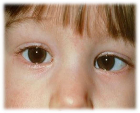
- WHY DOES STRABISMUS OCCUR ?
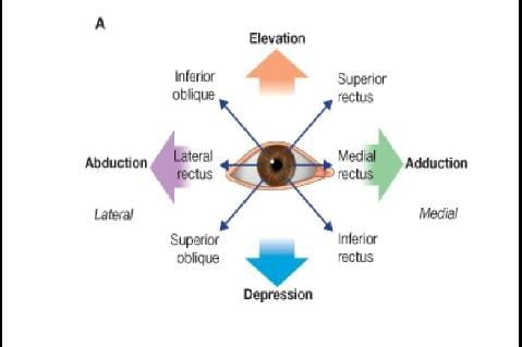
The position of each eyeball is maintained by six extraocular muscles. These muscles get their innervation from the 3rd, 4th and 6th cranial nerves (nerves that originate in the brain). These muscles move the eyes in various directions. Any weakness in movements either due to central mechanisms or due to direct involvement of the nerves or the mechanical restriction of eye movements can result in strabismus.
TYPES OF STRABISMUS :
Strabismus can be either latent or manifest. In latent squint,the tendency to squint is present, but kept in check by the fusion mechanisms of the eyes. Whenever fusion is interrupted, or when it is weak either due to illness or fatigue, the squint becomes manifest or apparent.
Manifest squint :
Manifest squint is of two types –
Comitant (degree of deviation constant in all directions of gaze and no limitation of eye movements)
Incomitant (deviation varies in different directions of gaze with limitation of eye movements)
PSEUDOSTABISMUS :
Pseudostrabismus is apparent squints. Pseudo means false, so pseudostrabismus means false squints. Normally, when both eyes are directed at a distant object the visual axes are parallel while the optical axes are slightly divergent. The “angle gamma” between the two is about 5 deg. and usually negligible , but in cases of high hypermetropia and myopia it may be considerable , giving rise to an apparent divergence in the former and convergence in the latter condition. Binocular vision is present & diplopia never occurs.
In pseudostabismus (apparent squint), the visual axes are in fact parallel, but the eye seem to have a squint :
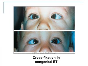
- Pseudoesotropia or apparent convergent squint may be associated with a prominent epicanthal fold (which covers the normally visible nasal aspect of the globe and gives false impression of esotropia) and negative angle kappa.
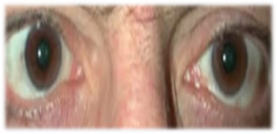
- Pseudoexotropia or apparent divergent squint may be associated with hypertelorism, a condition of wide separation of the two eyes, and positive angle kappa.
Causes :
Anatomical features
- Epicanthus :
- Skin fold , lead to apparent convergent squint (less white sclera is seen normally and the child “looks cross-eyed”).
- Mongolian face , or as congenital abnormality.
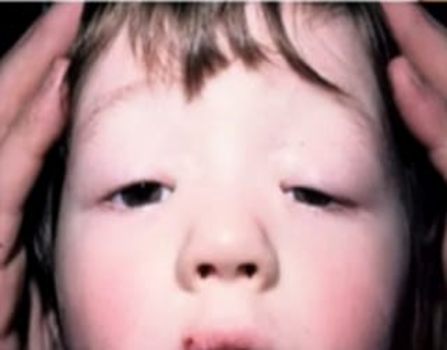
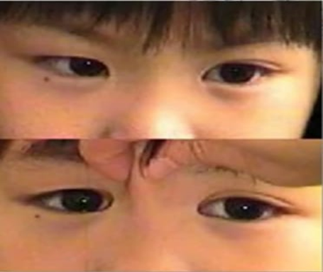
- Lateral eyelid adhesion (angular blepharitis complications) less white sclera is seen temporally and the patient “looks walked – eyed”).
- Abnormal inter – pupillary distance (IPD normally 60-64 mm)
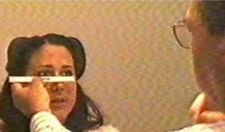
- Narrow IPD (apparent convergent squint).
- Wide IPD and wide nasal bridge (hyperterolism – apparent divergent squint).
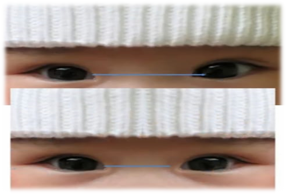
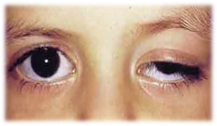
- Position of lid (vertical squint) :
- Ptosis
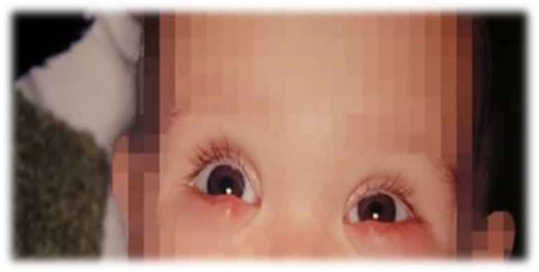
Passive edema of lower lid
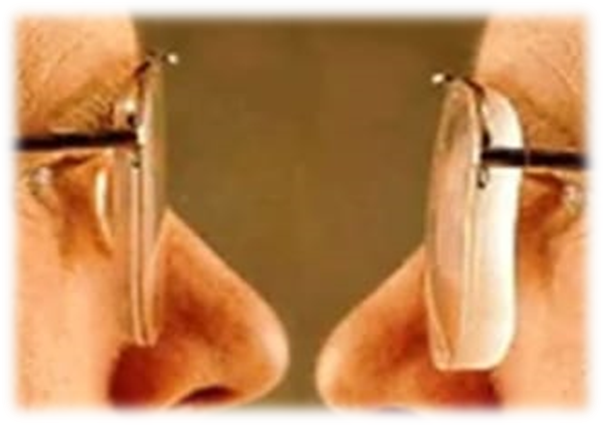
High errors of refraction :
- High myopia (apparent convergent squint-a negative angle alpha)
- High hyperopia (apparent divergent squint-a large positive angle alpha)
- Optic axis :
- Line which passes through center of curvature of the cornea and the lens and cuts the retina between the fovea and optic disc (nasal to central of the pupil)
- All the optical media of the eye are centered around it.
- Visual axis :
- Line which passes through object of regard , nodal point of the eye and fovea
- Cuts the cornea nasal to the optic axis and cuts the retina temporal to the optic axis.
- The two axes meet at nodal point ;
- Point where rays entering the eye undergo no refraction
- Present at the posterior pole of the crystalline lens.
- Corresponding to the optical center of a simple lens.
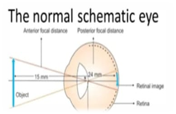
- Angle alpha ;
- Angle between the visual axis & the optic axis
- Normally the visual axis cut the cornea nasal to the optic axis & in this case , the angle is called positive angle alpha.
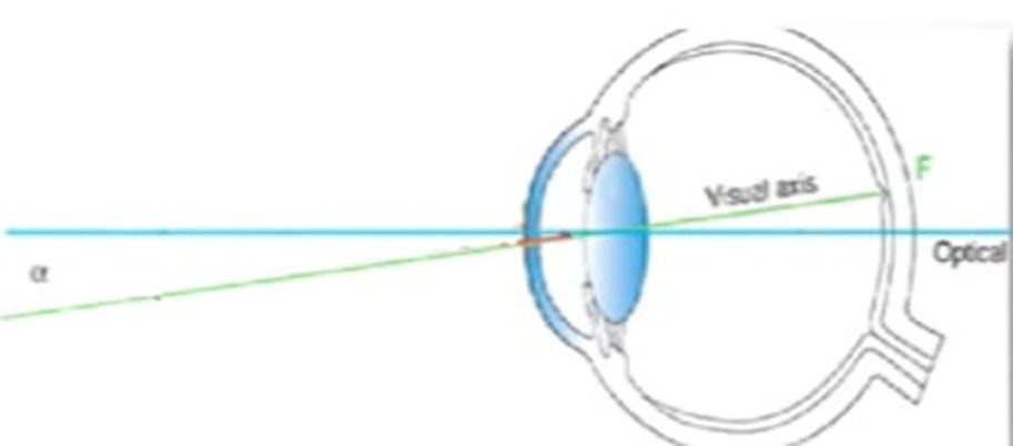
- Normally it is +5 degrees
- Angle alpha can be measured by major amblyoscope.
- Angle alpha (hyperopia) :
In high hypermetropia , angle alpha becomes large positive and in order to see , the optic axis must be displaced laterally to bring the visual axis forwards & then create apparent divergent squint.
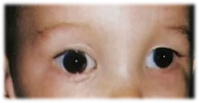
- Angle alpha (myopia) :
In high myopia due to enlargement of the posterior segment , the macula changes its position to cut its position. Then the visual axis will changes its position to cut the cornea temporal to the optic axis (angle alpha is called negative) & then create apparent convergent squint.
OBSERVATION :
A ‘Pseudo-Squint’ is sometimes found in a very young child in whom the centres of the cornea lie, comparatively speaking, nearer the inner walls of the orbits than they do later in life. Such a child on looking to the right appears to over converge with the left eye, and vice versa. Examination of the two light reflexes will show by their correspondence upon the cornea that no true squint is present.
TREATMENT :
- Glasses can normally correct the squint if hypermetropia or long-sightedness is causing it.
- Eye patch that is worn over the good eye can get the other (squint) eye to work better.
- Botulinum toxin injection or botox is injected into a muscle on the eye’s surface. Botox weakens the injected muscle for a short duration. Thus, it helps the eyes to align as it should be.
Eye drops and eye exercises too can help.
- Orthoptic treatment makes use of fusional training exercises and may be useful in some cases.
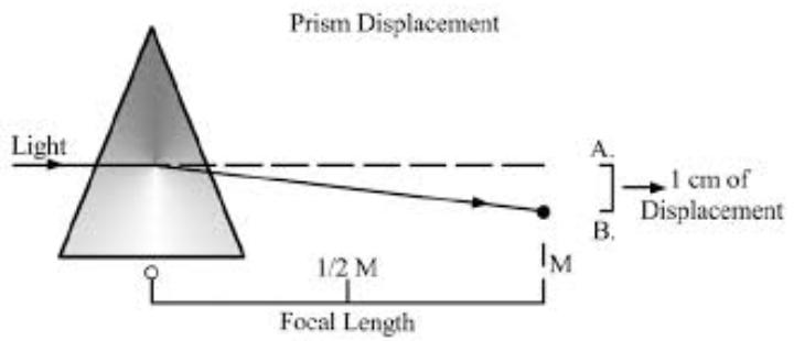
- Prism therapy : Prisms incorporated into spectacles are extremely helpful in improving the appearance of the eyes and facilitating the patient’s communication and interaction with others. It alleviates difficulties associated with misalignment. It is useful for older patients with limited fusional capabilities.
- Surgery is an option to go only when other treatments fail to be effective. It can restore binocular vision by realigning the eyes. The surgeon shifts the muscle that bends the eye to a new position. Sometimes the surgeon needs to operate both eyes to achieve the right balance.

